Approach
The clinical presentation of hepatitis D virus (HDV) infection ranges from asymptomatic to acute liver failure, cirrhosis, chronic liver disease, hepatic decompensation, and hepatocellular carcinoma.[1] Cirrhosis of the liver occurs in up to 70% of patients with chronic infection.[34] Presenting signs and symptoms depend on numerous factors, including the acuteness of infection and whether there is co-infection (i.e., simultaneous infection with hepatitis B virus [HBV]) or superinfection (i.e., HDV infection in a person already chronically infected with HBV).[16]
Co-infection leads to acute HDV and HBV infection, which is usually a self-limited illness but is more likely to lead to severe illness and acute liver failure (fulminant hepatitis) than is acute HBV infection.[17] Since HDV is dependent on HBV, the progression to chronic infection mirrors that of chronic hepatitis B, with younger age at infection predicting higher risk of chronicity.
Superinfection can present as a severe hepatitis, either in someone not known to have chronic HBV infection or as a flare of disease in someone with chronic HBV infection. Since the patient already has chronic HBV infection, progression to chronic HDV infection is almost universal.[35]
Evolution to chronic infection occurs in just 2% of patients who have co-infection compared with over 90% of patients who have superinfection.[34]
As patients with both acute and chronic forms of HDV infection are often asymptomatic, infection should be suspected in patients at increased risk.[2] Since HDV infection occurs in the setting of HBV infection, patients with risk factors for HBV infection (e.g., increased likelihood of sexual exposure, injection drug use, being born in a highly endemic region) are also at risk for HDV infection.
Patients who test positive for hepatitis B surface antigen (HBsAg) should be screened with an HDV antibody test; if this is found to be positive, testing for HDV RNA should be performed.[1] Diagnosis of exposure is confirmed by the presence of anti-HDV antibodies, and diagnosis of a current infection by the presence of HDV RNA.[1][12] Assays for anti-HDV antibody and HDV RNA level are increasingly available; the first international external quality control for HDV RNA quantification via the World Health Organization has led to improvements in HDV diagnostics.[2]
History
Ask about risk factors for HDV infection, which include:[10][11][12][24]
HBV infection
Being born in, living in, or travel to geographical regions where HBV/HDV are endemic (e.g., parts of western and mid-Africa, Mongolia, eastern Europe, and parts of the Mediterranean)
Increased likelihood of sexual exposure (e.g., multiple sexual partners, commercial sex workers)
Having sexual relations with a person infected with HDV
Men who have sex with men
Injection drug use
History of incarceration
Family history of HBV/HDV infection
Family history of hepatocellular carcinoma
Perinatal exposure in an infant born to an HDV-infected mother
Being a household contact of a person with HDV infection
Haemodialysis
Multiple blood transfusions
Being a healthcare worker
Acute HDV infection
Acute HDV infection occurs after an incubation period of 3-7 weeks.[12] It may present as a self-limited flu-like illness with non-specific clinical symptoms that may include fatigue, anorexia, nausea, lethargy, arthritis/arthralgia, fever, and abdominal pain.[12][34] In some patients this may be followed by jaundice, ongoing nausea and fatigue, dark urine, and pale stools.[34][36]
[Figure caption and citation for the preceding image starts]: A patient with jaundice, demonstrating scleral icterusCDC. Dr Thomas F. Sellers/Emory University; used with permission [Citation ends].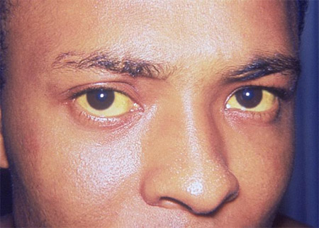
Presenting symptoms in acute HDV infection are indistinguishable from those associated with infection due to other forms of viral hepatitis, although clinical features tend to be more severe.[34] This is especially true in the case of injection drug users, where a high incidence of acute liver failure has been noted.[37]
Acute liver failure is a rare sequela of acute viral hepatitis; however, it occurs more frequently in acute HDV infection than in other forms of viral hepatitis infection.[34] Acute liver failure is defined by the onset of jaundice, coagulopathy (international normalised ratio [INR] >1.5), and hepatic encephalopathy in patients with no prior history of liver disease.[38][39][40] Abdominal pain, nausea, vomiting, and malaise are common symptoms, followed by jaundice, coagulopathy, and hepatic encephalopathy (confusion and changes in personality followed by somnolence and coma).[34] Right upper quadrant tenderness may also be found on examination.[34] More than half the cases of acute liver failure in HBsAg-positive patients are due to HDV infection rather than to HBV infection alone.[18] See Acute liver failure.
Chronic HDV infection
Chronic HDV infection is defined as presence of HDV beyond 6 months. Superinfection in the context of chronic HBV infection can lead to a flare of liver disease and is much more likely to become chronic in the majority (around 90%) of patients (unlike simultaneous HBV and HDV co-infection, which rarely becomes chronic).[18]
Although chronic HDV/HBV co-infection is often initially asymptomatic, it is the most severe type of chronic viral hepatitis and commonly leads to cirrhosis, liver failure, and hepatocellular carcinoma. Patients may therefore present with symptoms and signs of chronic liver disease and portal hypertension, such as fatigue, weight loss, spider naevi and palmar erythema. See Cirrhosis and Hepatocellular carcinoma.
Physical examination
Acute HDV infection
In patients with acute HDV infection, examination may reveal jaundice and, in some patients, right upper quadrant tenderness. Patients with acute HDV infection that has progressed to acute liver failure may also have evidence of encephalopathy (often with asterixis).
Chronic HDV infection
The physical examination in most patients with chronic HDV infection will be normal; however, patients with chronic HDV infection who have developed cirrhosis of the liver may have signs of chronic liver disease, including one or more of the following:
Palmar erythema
Spider naevi (in the distribution of the superior vena cava)
Scleral icterus
Ascites
Peripheral pitting oedema of the ankles/legs
Splenomegaly
Loss of secondary sexual characteristics, such as loss of secondary sexual hair and testicular atrophy in men
[Figure caption and citation for the preceding image starts]: Ascites in a female patient with cirrhosisScience Photo Library; used with permission [Citation ends].
Initial investigations
Given the importance of HDV infection to the long-term management of patients who are HBsAg-positive, all patients with confirmed hepatitis B infection should be tested for antibodies to hepatitis D (anti-HDV).[2]
Tests to characterise HBV infection
Perform serological tests to characterise HBV infection.[1] Serological markers include:
Hepatitis B surface antigen (HBsAg)
Antibody to hepatitis B surface antigen (anti-HBs)
Antibody to hepatitis B core antigen (anti-HBc) IgM and IgG
Hepatitis B e antigen (HBeAg)
Antibody to HBeAg (anti-HBe)
HBV DNA
A positive HBsAg establishes the diagnosis of HBV infection. It can be detected an average of 4 weeks (range 1-9 weeks) after virus exposure. Acute HBV infection is characterised by detection of HBsAg, HBeAg, HBV DNA, and anti-HBc IgM followed by anti-HBc IgG; a resolving acute infection is characterised by declining HBV DNA levels and disappearance of HBeAg with appearance of anti-HBe, followed by disappearance of HBsAg with appearance of anti-HBs. Anti-core IgM gradually disappears, while anti-core IgG (typically measured as 'total' antibody) remains detectable for life.
Chronic HBV infection is characterised by detection of HBsAg for more than 6 months. Anti-HBs are typically negative, while anti-HBc IgG are positive. Anti-HBc IgM may become transiently positive during flares. HBeAg and anti-HBe are used to determine the phase of chronic infection and may be positive or negative.
HBV DNA is essential for diagnosis and to determine the phase of infection; it may be undetectable or low in some patients with chronic HBV infection.[41][42]
Hepatitis B e-antigen (HBeAg) indicates high level viral replication and greater infectivity. The vast majority of patients with HBeAg-positive chronic HBV infection have biochemical and histological evidence of hepatitis; exceptions include children and young adults with perinatally acquired infection, who may show normal alanine aminotransferase (ALT) levels and normal liver histology. Absence of HBeAg does not necessarily indicate lack of virus replication as some patients have high or fluctuating HBV DNA levels and active hepatitis.
Consider testing for antibody to hepatitis B core antigen (anti-HBc) IgM, which may help distinguish individuals with HBV/HDV co-infection from HBsAg-positive individuals with HDV superinfection.[1]
Tests to characterise HDV infection
Perform serological tests for HDV.[1][12]
Serological markers include:
Antibody to hepatitis D (anti-HDV) IgM and IgG
HDV RNA
Note that:
Anti-HDV antibodies may persist after the clearance of HDV infection; therefore, testing for serum/plasma HDV RNA is required to confirm ongoing HDV infection.[1]
Serological tests are available to detect HDV antigen in serum, but they have variable performance and are not routinely recommended.
Anti-HDV IgM testing is not a reliable indicator of acute HDV infection as this test may become positive during chronic HDV infection.
HDV RNA assays are increasingly available, but require greater standardisation, particularly with some less common HDV genotypes.
Anti-HDV antibodies appear 3-4 weeks after HDV infection.[43] The result will be positive in people with chronic HDV infection, but levels can fluctuate based on the degree of replication.
The European Association for the Study of the Liver (EASL) recommends that screening for anti-HDV antibody should be performed with a validated assay at least once in all HBsAg-positive individuals, and that re-testing for anti-HDV antibody should be performed in those who initially test negative whenever clinically indicated (such as in those with aminotransferase flares or acute decompensation of chronic liver disease) and annually in those remaining at risk of infection.[1]
Guidelines from the American Association for the Study of Liver Diseases (AASLD) recommend risk-based screening of HBsAg-positive patients for HDV infection in the following situations:[2]
Elevated ALT or aspartate aminotransferase (AST) with low or undetectable HBV DNA
HIV infection
Hepatitis C virus (HCV) infection
History of injection drug use
Men who have sex with men
Those with multiple sexual partners or any history of sexually transmitted disease
Immigrants from areas with high HDV endemicity (western and mid-Africa, parts of the Mediterranean, Pacific Islands, Middle East, Mongolia, eastern Europe, and the Amazonian basin)[1]
In settings where universal HDV testing is not available, the World Health Organization (WHO) recommends prioritising testing in the following:[44]
People born in HDV-endemic regions
People with advanced liver disease, those receiving HBV antiviral therapy, and those with features that suggest HDV infection (e.g., low HBV DNA level with high ALT levels)
People considered to have an increased risk of HDV infection
Reflex testing (i.e., testing triggered automatically among all people who have a positive initial HBsAg screening test) may be used, where available.[44]
Tests to characterise liver disease status
Request the following in all patients with acute or chronic HDV infection:
Full blood count
Renal function tests
Coagulation profile
Liver biochemistries
AST
ALT
Total bilirubin (direct and indirect)
Alkaline phosphatase
Albumin
Check the patient's HIV (anti-HIV), hepatitis A (anti-HAV IgG/total), and hepatitis C (anti-HCV antibody ± HCV RNA) status, as this affects management options.
Perform abdominal imaging; ultrasound is a reasonable first test in all patients to evaluate the liver for advanced fibrosis, cirrhosis, portal hypertension, and for hepatocellular carcinoma.[41]
Triphasic contrast computed tomography or contrast magnetic resonance imaging are alternatives, particularly in patients with advanced fibrosis or cirrhosis.
Other investigations
Non-invasive methods of assessing liver fibrosis (e.g., transient elastography using magnetic resonance or FibroScan, fibrosis biomarkers) can be obtained to determine whether surveillance for hepatocellular carcinoma is required, but are seldom needed to assess therapy decisions.[1] Note that specific cut-off values for these tests are not well defined.[1]
Consider liver biopsy where results may contribute to the patient’s management, or for grading and staging liver disease when clinical signs of cirrhosis or evidence of cirrhosis (via imaging techniques) are not present.[1] Do not obtain liver biopsy routinely unless there is a diagnostic dilemma or if there is debate regarding whether treatment is required.[1]
Use of this content is subject to our disclaimer