Images and videos
Images


Otitis media with effusion
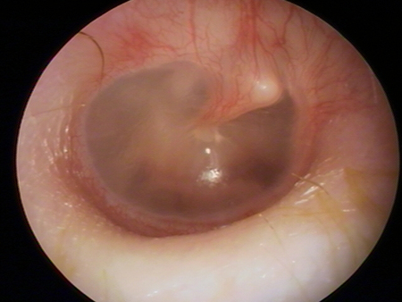
Otoscopy of otitis media with effusion, showing air fluid levels or bubbles, with normal tympanic membrane landmarks
From the personal collection of Dr Armengol
See this image in context in the following section/s:
Otitis media with effusion
Normal (type A) tympanogram
From the collection of Erica R. Thaler; used with permission
See this image in context in the following section/s:
Otitis media with effusion
Type B tympanogram; flat compliance curve demonstrates no movement of the tympanic membrane
From the collection of Erica R. Thaler; used with permission
See this image in context in the following section/s:
Otitis media with effusion
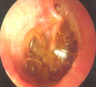
Appearance of tympanic membrane in otitis media with effusion, showing bubbles and serous fluid in the inferior aspect
Farboud A. BMJ. 2011;343:d3770
See this image in context in the following section/s:
Otitis media with effusion
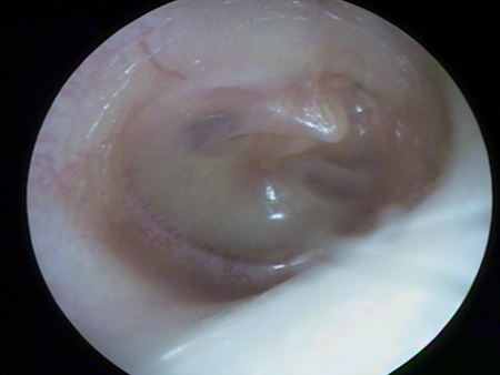
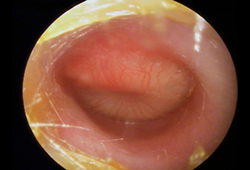
Otoscopy appearance of a bulging, erythematous tympanic membrane and absent landmarks
From the personal collection of Dr Armengol
See this image in context in the following section/s:
Otitis media with effusion
Otoscopy of myringitis, showing erythema and injection of the tympanic membrane in the neutral position
From the personal collection of Dr Armengol
See this image in context in the following section/s:
Otitis media with effusion
Type C tympanogram, demonstrating a malfunctioning Eustachian tube
From the collection of Erica R. Thaler; used with permission
See this image in context in the following section/s:
Use of this content is subject to our disclaimer