Otitis media with effusion (OME) is the presence of middle ear effusion with no signs and symptoms of acute infection (fever, ear pain, discharge from the ear, tympanic membrane bulging, and erythema).[2]Rosenfeld RM, Shin JJ, Schwartz SR, et al. Clinical practice guideline: otitis media with effusion (update). Otolaryngol Head Neck Surg. 2016 Feb;154(1 suppl):S1-41.
https://aao-hnsfjournals.onlinelibrary.wiley.com/doi/10.1177/0194599815623467
http://www.ncbi.nlm.nih.gov/pubmed/26832942?tool=bestpractice.com
It is predominantly a clinical diagnosis, consisting of symptoms and physical exam findings consistent with the presence of middle ear effusion (e.g., otoscopy/pneumatic otoscopy), and/or tympanometry.
OME may follow or precede acute otitis media (AOM) and the risk factors and presenting symptoms of both will overlap. However, it is important to differentiate OME from AOM because OME is not due to an acute infection and therefore should not be treated as such.[50]Lieberthal AS, Carroll AE, Chonmaitree T, et al. The diagnosis and management of acute otitis media. Pediatrics. 2013 Feb 25;131(3):e964-99.
https://pediatrics.aappublications.org/content/131/3/e964.long
http://www.ncbi.nlm.nih.gov/pubmed/23439909?tool=bestpractice.com
While middle ear effusion is present in both conditions (and is a hallmark of both), symptoms and signs of acute inflammation are not present in OME. See Acute otitis media.
History
The signs and symptoms that support a diagnosis of OME vary with the age of the patient. Both children and adults may even be asymptomatic or minimally symptomatic.[2]Rosenfeld RM, Shin JJ, Schwartz SR, et al. Clinical practice guideline: otitis media with effusion (update). Otolaryngol Head Neck Surg. 2016 Feb;154(1 suppl):S1-41.
https://aao-hnsfjournals.onlinelibrary.wiley.com/doi/10.1177/0194599815623467
http://www.ncbi.nlm.nih.gov/pubmed/26832942?tool=bestpractice.com
Older children and adults
In older children and in adults, symptoms typically consist of hearing loss, aural fullness or pressure, and a feeling of ear blockage.[51]Schilder AG, Bhutta MF, Butler CC, et al. Eustachian tube dysfunction: consensus statement on definition, types, clinical presentation and diagnosis. Clin Otolaryngol. 2015 Oct;40(5):407-11.
https://www.ncbi.nlm.nih.gov/pmc/articles/pmid/26347263
http://www.ncbi.nlm.nih.gov/pubmed/26347263?tool=bestpractice.com
Young children
In young children, symptoms may be more subtle, depending on the age and language development of the child, including poor balance, poor school performance, and behavioural problems.[2]Rosenfeld RM, Shin JJ, Schwartz SR, et al. Clinical practice guideline: otitis media with effusion (update). Otolaryngol Head Neck Surg. 2016 Feb;154(1 suppl):S1-41.
https://aao-hnsfjournals.onlinelibrary.wiley.com/doi/10.1177/0194599815623467
http://www.ncbi.nlm.nih.gov/pubmed/26832942?tool=bestpractice.com
Ask if the symptoms have had an impact on the child’s home life or at school.[52]Farboud A, Skinner R, Pratap R. Otitis media with effusion ("glue ear"). BMJ. 2011;343:d3770.
http://www.ncbi.nlm.nih.gov/pubmed/21727164?tool=bestpractice.com
Parents may notice signs of poor hearing, such as the child not responding when being called. Ask about indistinct speech, delayed language development or inattention. A parent or carer may also describe behavioural problems or slow progress within an education setting.[52]Farboud A, Skinner R, Pratap R. Otitis media with effusion ("glue ear"). BMJ. 2011;343:d3770.
http://www.ncbi.nlm.nih.gov/pubmed/21727164?tool=bestpractice.com
Pre-lingual children
Pre-lingual children may exhibit signs of aural discomfort such as tugging or scratching the pinna. They may also present with imbalance and impaired gross motor skills, as OME can affect the peripheral vestibular system.[3]Waldron MN, Matthews JN, Johnson IJ. The effect of otitis media with effusions on balance in children. Clin Otolaryngol Allied Sci. 2004 Aug;29(4):318-20.
http://www.ncbi.nlm.nih.gov/pubmed/15270815?tool=bestpractice.com
[4]Cohen H, Friedman EM, Lai D, et al. Balance in children with otitis media with effusion. Int J Pediatr Otorhinolaryngol. 1997 Dec 10;42(2):107-15.
http://www.ncbi.nlm.nih.gov/pubmed/9692620?tool=bestpractice.com
In infants, ask about the results of the newborn hearing screening. Infants and children may present after a failed hearing screen or during the work up of speech delay.
Chronic OME
Ask about the duration of symptoms. Chronic OME is effusion that is persistent for ≥3 months, either from the date of onset, if known, or from the date of first diagnosis of middle ear effusion.[53]Rosenfeld RM, Tunkel DE, Schwartz SR, et al. Clinical practice guideline: tympanostomy tubes in children (update). Otolaryngol Head Neck Surg. 2022 Feb;166(1_suppl):S1-55.
https://aao-hnsfjournals.onlinelibrary.wiley.com/doi/10.1177/01945998211065662
http://www.ncbi.nlm.nih.gov/pubmed/35138954?tool=bestpractice.com
[1]Simon F, Haggard M, Rosenfeld RM, et al. International consensus (ICON) on management of otitis media with effusion in children. Eur Ann Otorhinolaryngol Head Neck Dis. 2018 Feb;135(1s):S33-9.
https://www.sciencedirect.com/science/article/pii/S187972961830005X
http://www.ncbi.nlm.nih.gov/pubmed/29398506?tool=bestpractice.com
Risk factors
Patient and environmental factors can increase a child’s risk of OME. Risk factors for OME include:
Young age (i.e., 6 months to 4 years)[2]Rosenfeld RM, Shin JJ, Schwartz SR, et al. Clinical practice guideline: otitis media with effusion (update). Otolaryngol Head Neck Surg. 2016 Feb;154(1 suppl):S1-41.
https://aao-hnsfjournals.onlinelibrary.wiley.com/doi/10.1177/0194599815623467
http://www.ncbi.nlm.nih.gov/pubmed/26832942?tool=bestpractice.com
Recent or current upper respiratory tract infection[2]Rosenfeld RM, Shin JJ, Schwartz SR, et al. Clinical practice guideline: otitis media with effusion (update). Otolaryngol Head Neck Surg. 2016 Feb;154(1 suppl):S1-41.
https://aao-hnsfjournals.onlinelibrary.wiley.com/doi/10.1177/0194599815623467
http://www.ncbi.nlm.nih.gov/pubmed/26832942?tool=bestpractice.com
History of acute otitis media[2]Rosenfeld RM, Shin JJ, Schwartz SR, et al. Clinical practice guideline: otitis media with effusion (update). Otolaryngol Head Neck Surg. 2016 Feb;154(1 suppl):S1-41.
https://aao-hnsfjournals.onlinelibrary.wiley.com/doi/10.1177/0194599815623467
http://www.ncbi.nlm.nih.gov/pubmed/26832942?tool=bestpractice.com
Craniofacial anomalies[2]Rosenfeld RM, Shin JJ, Schwartz SR, et al. Clinical practice guideline: otitis media with effusion (update). Otolaryngol Head Neck Surg. 2016 Feb;154(1 suppl):S1-41.
https://aao-hnsfjournals.onlinelibrary.wiley.com/doi/10.1177/0194599815623467
http://www.ncbi.nlm.nih.gov/pubmed/26832942?tool=bestpractice.com
Eustachian tube dysfunction[41]Matsune S, Sando I, Takahashi H. Insertion of the tensor veli palatini muscle into the eustachian tube cartilage in cleft palate cases. Ann Otol Rhinol Laryngol. 1991 Jun;100(6):439-46.
http://www.ncbi.nlm.nih.gov/pubmed/2058982?tool=bestpractice.com
Genetic predisposition[2]Rosenfeld RM, Shin JJ, Schwartz SR, et al. Clinical practice guideline: otitis media with effusion (update). Otolaryngol Head Neck Surg. 2016 Feb;154(1 suppl):S1-41.
https://aao-hnsfjournals.onlinelibrary.wiley.com/doi/10.1177/0194599815623467
http://www.ncbi.nlm.nih.gov/pubmed/26832942?tool=bestpractice.com
[18]Casselbrant ML, Mandel EM, Fall PA, et al. The heritability of otitis media: a twin and triplet study. JAMA. 1999 Dec 8;282(22):2125-30.
https://jamanetwork.com/journals/jama/fullarticle/192180
http://www.ncbi.nlm.nih.gov/pubmed/10591333?tool=bestpractice.com
[1]Simon F, Haggard M, Rosenfeld RM, et al. International consensus (ICON) on management of otitis media with effusion in children. Eur Ann Otorhinolaryngol Head Neck Dis. 2018 Feb;135(1s):S33-9.
https://www.sciencedirect.com/science/article/pii/S187972961830005X
http://www.ncbi.nlm.nih.gov/pubmed/29398506?tool=bestpractice.com
Group childcare attendance[2]Rosenfeld RM, Shin JJ, Schwartz SR, et al. Clinical practice guideline: otitis media with effusion (update). Otolaryngol Head Neck Surg. 2016 Feb;154(1 suppl):S1-41.
https://aao-hnsfjournals.onlinelibrary.wiley.com/doi/10.1177/0194599815623467
http://www.ncbi.nlm.nih.gov/pubmed/26832942?tool=bestpractice.com
[27]Nguyen LH, Manoukian JJ, Yoskovitch A, et al. Adenoidectomy: selection criteria for surgical cases of otitis media. Laryngoscope. 2004 May;114(5):863-6.
http://www.ncbi.nlm.nih.gov/pubmed/15126745?tool=bestpractice.com
Allergy[1]Simon F, Haggard M, Rosenfeld RM, et al. International consensus (ICON) on management of otitis media with effusion in children. Eur Ann Otorhinolaryngol Head Neck Dis. 2018 Feb;135(1s):S33-9.
https://www.sciencedirect.com/science/article/pii/S187972961830005X
http://www.ncbi.nlm.nih.gov/pubmed/29398506?tool=bestpractice.com
Environmental tobacco smoke[2]Rosenfeld RM, Shin JJ, Schwartz SR, et al. Clinical practice guideline: otitis media with effusion (update). Otolaryngol Head Neck Surg. 2016 Feb;154(1 suppl):S1-41.
https://aao-hnsfjournals.onlinelibrary.wiley.com/doi/10.1177/0194599815623467
http://www.ncbi.nlm.nih.gov/pubmed/26832942?tool=bestpractice.com
History of sinonasal disease
Nasopharyngeal malignancy or other mass
Gastro-oesophageal reflux disease[1]Simon F, Haggard M, Rosenfeld RM, et al. International consensus (ICON) on management of otitis media with effusion in children. Eur Ann Otorhinolaryngol Head Neck Dis. 2018 Feb;135(1s):S33-9.
https://www.sciencedirect.com/science/article/pii/S187972961830005X
http://www.ncbi.nlm.nih.gov/pubmed/29398506?tool=bestpractice.com
Low socioeconomic status.[10]Zhang Y, Xu M, Zhang J, et al. Risk factors for chronic and recurrent otitis media-a meta-analysis. PLoS One. 2014;9(1):e86397.
https://www.ncbi.nlm.nih.gov/pmc/articles/PMC3900534
http://www.ncbi.nlm.nih.gov/pubmed/24466073?tool=bestpractice.com
Physical examination
Examine the patient’s upper respiratory tract and ears. Note any aural discharge. Persistent discharge, along with atypical otoscopy, may indicate a cholesteatoma, which requires urgent referral for specialist assessment of the ear, nose, and throat.[52]Farboud A, Skinner R, Pratap R. Otitis media with effusion ("glue ear"). BMJ. 2011;343:d3770.
http://www.ncbi.nlm.nih.gov/pubmed/21727164?tool=bestpractice.com
Examine both ears with an otoscope. The tympanic membrane may appear dark, with an amber, grey or blue hue. There may be bubbles or an air-fluid level.[2]Rosenfeld RM, Shin JJ, Schwartz SR, et al. Clinical practice guideline: otitis media with effusion (update). Otolaryngol Head Neck Surg. 2016 Feb;154(1 suppl):S1-41.
https://aao-hnsfjournals.onlinelibrary.wiley.com/doi/10.1177/0194599815623467
http://www.ncbi.nlm.nih.gov/pubmed/26832942?tool=bestpractice.com
In chronic OME (i.e. ≥3 months’ duration) the tympanic membrane may be retracted, in which case normal landmarks such as the short process of the malleus will appear more prominent.[50]Lieberthal AS, Carroll AE, Chonmaitree T, et al. The diagnosis and management of acute otitis media. Pediatrics. 2013 Feb 25;131(3):e964-99.
https://pediatrics.aappublications.org/content/131/3/e964.long
http://www.ncbi.nlm.nih.gov/pubmed/23439909?tool=bestpractice.com
However, a normal looking tympanic membrane does not exclude OME unless pneumatic otoscopy is also normal.
[Figure caption and citation for the preceding image starts]: Otoscopy of otitis media with effusion, showing air fluid levels or bubbles, with normal tympanic membrane landmarksFrom the personal collection of Dr Armengol [Citation ends].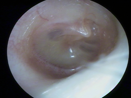 [Figure caption and citation for the preceding image starts]: Appearance of tympanic membrane in otitis media with effusion, showing bubbles and serous fluid in the inferior aspectFarboud A. BMJ. 2011;343:d3770 [Citation ends].
Initial investigations
OME is predominantly a clinical diagnosis. Pneumatic otoscopy should be performed, where possible, to confirm the presence of a middle ear effusion. Objective testing with tympanometry may be appropriate in certain patients for whom the diagnosis remains uncertain after the physical examination and pneumatic otoscopy. Audiological evaluation is needed to assess hearing loss in patients with chronic OME, or for OME of any duration in a child at risk of developmental sequelae (see Audiology section below).[2]Rosenfeld RM, Shin JJ, Schwartz SR, et al. Clinical practice guideline: otitis media with effusion (update). Otolaryngol Head Neck Surg. 2016 Feb;154(1 suppl):S1-41.
https://aao-hnsfjournals.onlinelibrary.wiley.com/doi/10.1177/0194599815623467
http://www.ncbi.nlm.nih.gov/pubmed/26832942?tool=bestpractice.com
Pneumatic otoscopy
Where possible, use pneumatic otoscopy to make the diagnosis in a patient with symptoms or signs suggestive of OME.[2]Rosenfeld RM, Shin JJ, Schwartz SR, et al. Clinical practice guideline: otitis media with effusion (update). Otolaryngol Head Neck Surg. 2016 Feb;154(1 suppl):S1-41.
https://aao-hnsfjournals.onlinelibrary.wiley.com/doi/10.1177/0194599815623467
http://www.ncbi.nlm.nih.gov/pubmed/26832942?tool=bestpractice.com
[1]Simon F, Haggard M, Rosenfeld RM, et al. International consensus (ICON) on management of otitis media with effusion in children. Eur Ann Otorhinolaryngol Head Neck Dis. 2018 Feb;135(1s):S33-9.
https://www.sciencedirect.com/science/article/pii/S187972961830005X
http://www.ncbi.nlm.nih.gov/pubmed/29398506?tool=bestpractice.com
The value of pneumatic otoscopy over standard otoscopy is that it can detect reduced tympanic mobility, which may be the only sign of effusion in a patient with OME.[2]Rosenfeld RM, Shin JJ, Schwartz SR, et al. Clinical practice guideline: otitis media with effusion (update). Otolaryngol Head Neck Surg. 2016 Feb;154(1 suppl):S1-41.
https://aao-hnsfjournals.onlinelibrary.wiley.com/doi/10.1177/0194599815623467
http://www.ncbi.nlm.nih.gov/pubmed/26832942?tool=bestpractice.com
It has a sensitivity and specificity of 94% and 80%, respectively, when compared to myringotomy.[54]Shekelle P, Takata G, Chan LS, et al. Diagnosis, natural history, and late effects of otitis media with effusion. Evid Rep Technol Assess (Summ). 2002 Jun;(55):1-5.
https://www.ncbi.nlm.nih.gov/pmc/articles/PMC4781261
http://www.ncbi.nlm.nih.gov/pubmed/12945555?tool=bestpractice.com
Make a diagnosis of OME based on reduced or restricted movement of the tympanic membrane; a complete absence of movement is not needed to be diagnostic.[2]Rosenfeld RM, Shin JJ, Schwartz SR, et al. Clinical practice guideline: otitis media with effusion (update). Otolaryngol Head Neck Surg. 2016 Feb;154(1 suppl):S1-41.
https://aao-hnsfjournals.onlinelibrary.wiley.com/doi/10.1177/0194599815623467
http://www.ncbi.nlm.nih.gov/pubmed/26832942?tool=bestpractice.com
Pneumatic otoscopy might be difficult to perform in young children, in part due to their narrow ear canals and also due to the tendency of young children to move as the investigation is being performed.[55]Venekamp RP, Schilder AGM, van den Heuvel M, et al. Acute otitis media in children. BMJ. 2020 Nov 18;371:m4238.
http://www.ncbi.nlm.nih.gov/pubmed/33208317?tool=bestpractice.com
Tympanometry
For patients who are difficult to examine and for patients in whom the diagnosis is uncertain following pneumatic otoscopy, perform tympanometry, where possible, to provide objective information on tympanic membrane mobility and middle ear compliance.[2]Rosenfeld RM, Shin JJ, Schwartz SR, et al. Clinical practice guideline: otitis media with effusion (update). Otolaryngol Head Neck Surg. 2016 Feb;154(1 suppl):S1-41.
https://aao-hnsfjournals.onlinelibrary.wiley.com/doi/10.1177/0194599815623467
http://www.ncbi.nlm.nih.gov/pubmed/26832942?tool=bestpractice.com
Tympanometric tracings (or curves) are classified into three main types:[2]Rosenfeld RM, Shin JJ, Schwartz SR, et al. Clinical practice guideline: otitis media with effusion (update). Otolaryngol Head Neck Surg. 2016 Feb;154(1 suppl):S1-41.
https://aao-hnsfjournals.onlinelibrary.wiley.com/doi/10.1177/0194599815623467
http://www.ncbi.nlm.nih.gov/pubmed/26832942?tool=bestpractice.com
[56]Onusko E. Tympanometry. Am Fam Physician. 2004 Nov 1;70(9):1713-20.
https://www.aafp.org/pubs/afp/issues/2004/1101/p1713.html
http://www.ncbi.nlm.nih.gov/pubmed/15554489?tool=bestpractice.com
Type A indicates a normal ear with a low probability of effusion
Type B is the typical result to confirm OME (but a type B trace can also be seen with other conditions)
Type C suggests negative pressure, which may be related to OME. It should be correlated with other findings to confirm a diagnosis of OME.
[Figure caption and citation for the preceding image starts]: Normal (type A) tympanogramFrom the collection of Erica R. Thaler; used with permission [Citation ends]. [Figure caption and citation for the preceding image starts]: Type B tympanogram; flat compliance curve demonstrates no movement of the tympanic membraneFrom the collection of Erica R. Thaler; used with permission [Citation ends].
 [Figure caption and citation for the preceding image starts]: Type C tympanogram, demonstrating a malfunctioning Eustachian tubeFrom the collection of Erica R. Thaler; used with permission [Citation ends].
Audiology
In patients with chronic OME, or for OME of any duration in an at-risk child (see list below), perform an audiogram to establish baseline hearing levels and the impact of OME on the patient’s hearing. OME can cause no hearing loss through to a moderate conductive hearing loss.[2]Rosenfeld RM, Shin JJ, Schwartz SR, et al. Clinical practice guideline: otitis media with effusion (update). Otolaryngol Head Neck Surg. 2016 Feb;154(1 suppl):S1-41.
https://aao-hnsfjournals.onlinelibrary.wiley.com/doi/10.1177/0194599815623467
http://www.ncbi.nlm.nih.gov/pubmed/26832942?tool=bestpractice.com
Children who have the following conditions are considered to be at risk of developmental sequelae as a result of OME:[53]Rosenfeld RM, Tunkel DE, Schwartz SR, et al. Clinical practice guideline: tympanostomy tubes in children (update). Otolaryngol Head Neck Surg. 2022 Feb;166(1_suppl):S1-55.
https://aao-hnsfjournals.onlinelibrary.wiley.com/doi/10.1177/01945998211065662
http://www.ncbi.nlm.nih.gov/pubmed/35138954?tool=bestpractice.com
Permanent non-OME-related hearing loss
Speech and language delay or disorder
Autism-spectrum disorder
Genetic syndromes or craniofacial disorders associated with cognitive, speech, or language delays
Blindness or uncorrectable visual impairment
Cleft palate
Developmental delay
Intellectual disability, learning disorders, or attention deficit/hyperactivity disorder.
Hearing loss can lead to speech delay, behavioural problems, and poor school performance in children, so it is important to identify. A hearing test should be performed that is appropriate for the patient’s age.[2]Rosenfeld RM, Shin JJ, Schwartz SR, et al. Clinical practice guideline: otitis media with effusion (update). Otolaryngol Head Neck Surg. 2016 Feb;154(1 suppl):S1-41.
https://aao-hnsfjournals.onlinelibrary.wiley.com/doi/10.1177/0194599815623467
http://www.ncbi.nlm.nih.gov/pubmed/26832942?tool=bestpractice.com
[1]Simon F, Haggard M, Rosenfeld RM, et al. International consensus (ICON) on management of otitis media with effusion in children. Eur Ann Otorhinolaryngol Head Neck Dis. 2018 Feb;135(1s):S33-9.
https://www.sciencedirect.com/science/article/pii/S187972961830005X
http://www.ncbi.nlm.nih.gov/pubmed/29398506?tool=bestpractice.com
Tests may include pure tone audiometry, speech audiometry, and bone-conduction.[1]Simon F, Haggard M, Rosenfeld RM, et al. International consensus (ICON) on management of otitis media with effusion in children. Eur Ann Otorhinolaryngol Head Neck Dis. 2018 Feb;135(1s):S33-9.
https://www.sciencedirect.com/science/article/pii/S187972961830005X
http://www.ncbi.nlm.nih.gov/pubmed/29398506?tool=bestpractice.com
The pure tone threshold average at 500, 1000, 2000, and 4000 Hz may show hearing loss, typically of around 28 dB, but can be up to 50 dB in those with OME.[57]Roberts J, Hunter L, Gravel J, et al. Otitis media, hearing loss, and language learning: controversies and current research. J Dev Behav Pediatr. 2004 Apr;25(2):110-22.
http://www.ncbi.nlm.nih.gov/pubmed/15083134?tool=bestpractice.com
[58]Dougherty W, Kesser BW. Management of conductive hearing loss in children. Otolaryngol Clin North Am. 2015 Dec;48(6):955-74.
http://www.ncbi.nlm.nih.gov/pubmed/26360369?tool=bestpractice.com
Consider any concerns from the parent or carer reporting the hearing loss. Severe hearing loss may indicate pathology other than or in addition to OME, such as congenital sensorineural hearing loss (genetic or non-genetic), inner ear malformations, and ossicular chain abnormalities, and should be appropriately investigated.
Developmental abnormalities or behavioural problems may affect the results of a routine hearing test, in which case referral to an otolaryngologist or audiologist may be required.[2]Rosenfeld RM, Shin JJ, Schwartz SR, et al. Clinical practice guideline: otitis media with effusion (update). Otolaryngol Head Neck Surg. 2016 Feb;154(1 suppl):S1-41.
https://aao-hnsfjournals.onlinelibrary.wiley.com/doi/10.1177/0194599815623467
http://www.ncbi.nlm.nih.gov/pubmed/26832942?tool=bestpractice.com
In infants with OME who fail a newborn hearing screen, follow-up is important to ensure that hearing is in fact normal once the OME resolves.[2]Rosenfeld RM, Shin JJ, Schwartz SR, et al. Clinical practice guideline: otitis media with effusion (update). Otolaryngol Head Neck Surg. 2016 Feb;154(1 suppl):S1-41.
https://aao-hnsfjournals.onlinelibrary.wiley.com/doi/10.1177/0194599815623467
http://www.ncbi.nlm.nih.gov/pubmed/26832942?tool=bestpractice.com
The results of hearing tests may influence the management plan.
Other investigations
In patients with chronic or recurrent OME, refer to otolaryngology for consideration of nasal endoscopy to assess for signs of chronic rhinosinusitis, as chronic rhinosinusitis with and without nasal polyposis has been associated with an increased risk of OME.[24]Finkelstein Y, Ophir D, Talmi YP, et al. Adult-onset otitis media with effusion. Arch Otolaryngol Head Neck Surg. 1994 May;120(5):517-27.
http://www.ncbi.nlm.nih.gov/pubmed/8172703?tool=bestpractice.com
[26]Parietti-Winkler C, Baumann C, Gallet P, et al. Otitis media with effusion as a marker of the inflammatory process associated to nasal polyposis. Rhinology. 2009 Dec;47(4):396-9.
http://www.ncbi.nlm.nih.gov/pubmed/19936366?tool=bestpractice.com
As a secondary investigation, nasopharyngeal endoscopy may be helpful in children for whom there is concern about concurrent nasal obstruction or prolonged unilateral OME to identify adenoid hypertrophy (providing that they are tolerant of the examination). Consider this more routinely in areas of high HIV prevalence due to the associated increased risk of nasopharyngeal anomalies, such as lymphoma.[1]Simon F, Haggard M, Rosenfeld RM, et al. International consensus (ICON) on management of otitis media with effusion in children. Eur Ann Otorhinolaryngol Head Neck Dis. 2018 Feb;135(1s):S33-9.
https://www.sciencedirect.com/science/article/pii/S187972961830005X
http://www.ncbi.nlm.nih.gov/pubmed/29398506?tool=bestpractice.com
In adults, refer to otolaryngology for consideration of nasopharyngeal endoscopy to assess the area by the torus tubarius (fossa of Rosenmueller), particularly if the effusion is unilateral.[24]Finkelstein Y, Ophir D, Talmi YP, et al. Adult-onset otitis media with effusion. Arch Otolaryngol Head Neck Surg. 1994 May;120(5):517-27.
http://www.ncbi.nlm.nih.gov/pubmed/8172703?tool=bestpractice.com
[59]Neel HB 3rd. Nasopharyngeal carcinoma: diagnosis, staging, and management. Oncology (Williston Park). 1992 Feb;6(2):87-102.
http://www.ncbi.nlm.nih.gov/pubmed/1532503?tool=bestpractice.com
Look for signs of nasopharyngeal carcinoma on nasal endoscopy as it is associated with adult-onset OME. The rate of nasopharyngeal carcinoma in adults with OME is around 5% to 6%.[28]Ho KY, Lee KW, Chai CY, et al. Early recognition of nasopharyngeal cancer in adults with only otitis media with effusion. J Otolaryngol Head Neck Surg. 2008 Jun;37(3):362-5.
http://www.ncbi.nlm.nih.gov/pubmed/19128640?tool=bestpractice.com
[24]Finkelstein Y, Ophir D, Talmi YP, et al. Adult-onset otitis media with effusion. Arch Otolaryngol Head Neck Surg. 1994 May;120(5):517-27.
http://www.ncbi.nlm.nih.gov/pubmed/8172703?tool=bestpractice.com