History and exam
Key diagnostic factors
common
uncommon
open fracture
Laceration or wound to skin/soft tissue with exposed fractured bone or visible bone fragments is suggestive of skull fracture.
palpable discrepancy in bone contour
Depressed or comminuted fractures are usually easily palpable. However, palpable changes in the bony cortex contour (step-offs) or palpable fracture fragments are rare in simple linear fractures.
Battle's sign
Blood pooling from basilar skull fractures can result in ecchymosis overlying the mastoid process. If present, it has a positive predictive value of 66% for basilar fractures.[1]
Most often associated with fractures of the petrous portion of the temporal bone.[1]
[Figure caption and citation for the preceding image starts]: Battle's sign: superficial ecchymosis over the mastoid processGert W van Dijk Pract Neurol 2011;11:50-55. [Citation ends].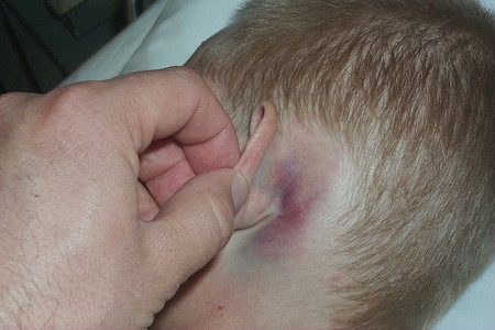
periorbital ecchymosis
bloody otorrhoea
cerebrospinal fluid rhinorrhoea
Most often associated with fractures of the anterior cranial fossa.[1]
Confirmation requires beta-2 transferrin test.
facial paralysis, nystagmus, or paraesthesia
Consequence of cranial nerve injury causing facial paralysis or nystagmus (VII) or paraesthesia (V); associated with basilar skull fractures.
Other diagnostic factors
common
evidence of trauma
Includes soft-tissue swelling, haematomas, crepitus, lacerations, and tenderness.
Not specific for skull fracture, although the presence of cranial haematomas is more suggestive of a skull fracture in children than in adults.[57] Absence of these features does not exclude fracture.
uncommon
cranial pain or headache
Non-specific symptom. Occipital skull fractures, crossing the transverse venous sinus, can cause venous sinus thrombosis, which in turn will cause raised intracranial pressure. In an awake and alert patient, this may present as intractable headaches (with nausea and vomiting).[30]
nausea/vomiting
Non-specific symptoms. Less typical in adults, occur much more frequently in adolescents and children. Occipital skull fractures, crossing the transverse venous sinus, can cause venous sinus thrombosis, which in turn will cause raised intracranial pressure. In an awake and alert patient, this may present as intractable headaches with nausea and vomiting.[30]
If the patient is a child, take a good history from the parent(s) regarding any vomiting since the head injury. A vomit is defined as a single discrete episode of vomiting; it is very common in younger children, and on its own is not of concern. When vomiting is the only symptom of head injury in a child, traumatic brain injury on CT is uncommon and clinically important traumatic brain injury is very uncommon.[58] Traumatic brain injury is more frequent in children when vomiting is accompanied by other signs or symptoms suggestive of traumatic brain injury.[58] However, request an urgent (i.e., within 1 hour) CT scan for any children with ≥3 discrete episodes of vomiting together with any one or more of the following risk factors:[28]
Witnessed loss of consciousness lasting more than 5 minutes
Abnormal drowsiness
A dangerous mechanism of injury (high-speed road traffic accident either as pedestrian, cyclist, or vehicle occupant, fall from a height of more than 3 m, high-speed injury from a projectile or other object)
Amnesia (antegrade or retrograde) lasting more than 5 minutes
Any current bleeding or clotting disorder (liver failure, haemophilia, taking anticoagulants or antiplatelets).
altered mental state/loss of consciousness
Loss of consciousness is most commonly related to underlying associated intracranial injury and is rare in isolated skull fractures.
The patient's neurological status should be assessed at initial presentation and subsequently monitored to help guide management decisions. Use the Glasgow Coma Scale (GCS) to assess any associated traumatic brain injury.[28] [ Glasgow Coma Scale Opens in new window ] Use the paediatric version in children <1 year.[28] Glasgow Coma Scale with modification for children Opens in new window In practice, the paediatric GCS is used until age 18-24 months since the standard GCS requires assessment of a verbal response as 'orientated' which is typically not possible in younger children and infants. Also use the GCS/paediatric GCS for subsequent monitoring to help guide management decisions.
abnormal pupillary reflexes
If present, can suggest herniation or brainstem injury.
Practical tip
If the patient’s pupils are dilated, take a thorough drug history. Be sure to include illicit drugs as well as medication (either taken by the patient independently or given prehospital, e.g., by the ambulance crew) which can cause dilation.
hearing loss
Basilar skull fractures affecting the mastoid process or the inner ear may present with facial nerve palsy or hearing loss due to the proximity of these structures. Conductive hearing loss may also present early (<3 weeks) because of haemotympanum with temporal bone fractures, or later (>6 weeks) with longitudinal temporal bone fracture with disruption of ossicular chain.
Use of this content is subject to our disclaimer