Recommendations
Urgent
Suspect cholangitis in patients with known risk factors and typical findings such as a history of pale stools and abdominal pain, and jaundice, fever, and right upper quadrant (RUQ) tenderness on examination.
Cholangitis can quickly become an acute, septic, life-threatening infection if not identified and treated promptly. Think 'Could this be sepsis?' based on acute deterioration in an adult patient in whom there is clinical evidence or strong suspicion of infection.[22][23][24] See Sepsis in adults.
The patient may present with non-specific or non-localised symptoms (e.g., acutely unwell with a normal temperature) or there may be severe signs with evidence of multi-organ dysfunction and shock.[22][23][24]
Remember that sepsis represents the severe, life-threatening end of infection.[25]
Use a systematic approach (e.g., National Early Warning Score 2 [NEWS2]), alongside your clinical judgement, to assess the risk of deterioration due to sepsis.[22][23][26][27] Consult local guidelines for the recommended approach at your institution.
Arrange urgent review by a senior clinical decision-maker (e.g., ST4 level doctor in the UK) if you suspect sepsis:[28]
Within 30 minutes for a patient who is critically ill (e.g., NEWS2 score of 7 or more, evidence of septic shock, or other significant clinical concerns)
Within 1 hour for a patient who is severely ill (e.g., NEWS2 score of 5 or 6).
Follow your local protocol for investigation and treatment of all patients with suspected sepsis, or those at risk. Start treatment promptly. Determine urgency of treatment according to likelihood of infection and severity of illness, or according to your local protocol.[27][28]
In the community: refer for emergency medical care in hospital (usually by blue-light ambulance in the UK) any patient who is acutely ill with a suspected infection and is:[24]
Deemed to be at high risk of deterioration due to organ dysfunction (as measured by risk stratification)
At risk of neutropenic sepsis.
Suspect severe disease if your patient is hypotensive or has mental status changes.[1][29]
Arrange for a transabdominal ultrasound as the initial diagnostic imaging investigation of choice.[30][31]
Order this test urgently if there are signs of severe cholangitis or the patient is high risk for sepsis.
Arrange subsequent abdominal CT scan with intravenous contrast if the ultrasound is inconclusive.
Abdominal CT scan is often done in patients with sepsis to rule out other causes of sepsis.
Key Recommendations
If you suspect sepsis, see Urgent recommendations and Sepsis in adults.
Suspect possible cholangitis if there are typical features in the patient’s history and physical examination, including:
Presence of risk factors (e.g., age >50 years, known HIV infection, past history of gallstones, or ERCP)
History of pale stools, upper abdominal pain
Fever, jaundice, and RUQ tenderness on examination (Charcot’s triad).
Take initial blood tests, including:
FBC
Coagulation profile
CRP
LFTs
Urea and electrolytes (including creatinine and magnesium)
Blood culture.[32]
Take an arterial blood gas sample, including lactate, if sepsis is suspected.
Arrange for a transabdominal ultrasound as the first imaging investigation.[30][31]
Consider an abdominal CT scan with intravenous contrast if the ultrasound is negative but the diagnosis is still suspected.
Consider magnetic resonance cholangiopancreatography (MRCP) or endoscopic ultrasound (depending on availability) before definitive treatment.[31]
Seek senior advice on which method of biliary decompression is most appropriate for your patient. These procedures may be diagnostic and therapeutic.
All patients with cholangitis eventually need biliary decompression, most commonly using endoscopic retrograde cholangiopancreatography (ERCP).[33]
Arrange early ERCP if there is a history of predisposing factors such as known biliary disease, or indwelling biliary prosthesis.
Other methods of biliary decompression include:
Percutaneous trans-hepatic cholangiography.
Acute cholangitis requires prompt diagnosis and treatment. Cholangitis can quickly become an acute, septic, life-threatening infection if not identified and treated promptly.[1] If you suspect sepsis, see Urgent recommendations and Sepsis in adults.
Diagnosis of acute cholangitis is based on typical history and laboratory findings and confirmed by imaging, endoscopic retrograde cholangiopancreatography (ERCP), or percutaneous biliary drainage in patients who cannot undergo ERCP.[1] The Tokyo guidelines provide a concise overview of the factors to be considered in the diagnosis and severity assessment of these patients.[1] Most patients will present with fever, jaundice, and right upper quadrant (RUQ) pain (Charcot's triad), although some patients with a significant infection may have a surprisingly benign appearance overall.[3] Patients with cholangitis typically have diffuse RUQ pain and not classic Murphy's sign.
Be aware that most patients will present with fever, jaundice, and right upper quadrant (RUQ) pain (Charcot's triad), and are typically aged over 50 years.
Take a detailed history:
Establish the presence of risk factors for acute cholangitis, with a focus on:[10]
Known gallstones
Prior endoscopic retrograde cholangiopancreatography (ERCP)
Surgical or radiological biliary tree intervention
HIV infection.
Determine if there is a current history of pale stools, pruritus, and jaundice.
Fever and RUQ pain are present in 65% to 90% of patients, though fever can be absent in older patients (those over 60 years old); 65% of patients will have RUQ tenderness.[1]
Putty/clay coloured stools may be seen due to deficient bile secretion to the small intestine.
Sensation of itch associated with any liver disease may be reported.
Jaundice is present in 60% to 70% of patients.[1] Hypotension is present in 30% of patients.[1]
Mental status changes occur in about 15% of patients.[1]
Hypotension and mental status changes are indicative of severe disease and are associated with a poor prognosis.[1][29]
Request the following blood tests in patients with suspected or known acute cholangitis (listed with common findings):
FBC: white blood cell count is typically >10.0 × 10⁹/L (>10,000/microlitre) (reference range 4.8 to 10.8 × 10⁹/L or 4800-10,800/microlitre).
CRP (a marker of inflammation): can be raised.
LFTs: hyperbilirubinaemia is almost always present and, if absent, cholangitis is less likely to be the true diagnosis. Abnormal liver function tests are usual findings, with raised serum alkaline phosphatase and transaminases being typical.
Urea and creatinine: raised renal function parameters are more common in severe disease states.
Electrolytes plus magnesium: possible decreases of serum potassium and magnesium.
Blood culture: positive blood culture rates among patients with acute cholangitis range from 21% to 71%.[32] Bacteria are usually gram-negative, but gram-positive bacteria and anaerobes are also implicated in cholangitis.[32]
Coagulation profile: abnormalities can include decreased platelets and raised prothrombin time.
ABG analysis and lactate (additional if there is concern about sepsis): metabolic acidosis is common in severe disease states and raised lactate is associated with sepsis.

How to take a venous blood sample from the antecubital fossa using a vacuum needle.
Perform transabdominal ultrasound in all patients presenting with right upper quadrant (RUQ) pain and suspected cholangitis.[30][31]
The main value of a transabdominal ultrasound is in detecting cholecystitis, which can mimic cholangitis, and to provide a limited evaluation of the biliary tree.
Transabdominal ultrasound is a quick, easy, and inexpensive initial diagnostic imaging modality. Its accuracy for detecting common bile duct (CBD) dilation is >90%, although the diameter of the CBD becomes a less useful parameter in patients who have previously undergone cholecystectomy, as physiological dilation of the CBD can occur in this setting.
Transabdominal ultrasound has a poor sensitivity for detecting mid to distal CBD stones.[1]
Consider abdominal computed tomography (CT) scan with intravenous contrast if there is high clinical suspicion of cholangitis, and ultrasound is negative.
Abdominal CT provides better anatomical detail of the biliary tree than transabdominal ultrasound. It is particularly useful to visualise the distal part of the CBD.
Abdominal CT is a better initial imaging choice than transabdominal ultrasound if neoplasm is suspected as the cause of cholangitis.
CT scans are contraindicated in patients with intravenous contrast dye allergy and may be detrimental to those with renal dysfunction.
Consider early endoscopic retrograde cholangiopancreatography (ERCP) for rapid diagnosis and therapy in patients with a history of biliary disease, an indwelling biliary prosthesis, or other predisposing factors.
[Figure caption and citation for the preceding image starts]: Endoscopic retrograde cholangiopancreatography reveals a large common bile duct (CBD) stone (arrow) in the mid-common bile ductFrom the collection of Douglas G. Adler; used with permission [Citation ends].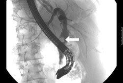 [Figure caption and citation for the preceding image starts]: Endoscopic photo of same patient following removal of large common bile duct (CBD) stone; note copious pus draining through the ampullaFrom the collection of Douglas G. Adler; used with permission [Citation ends].
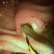
Percutaneous trans-hepatic cholangiography can provide direct evaluation of the biliary tree if ERCP is not available or is impossible to perform (e.g., post-Roux-en-Y gastric bypass; presence of oesophageal, pyloric, or duodenal stricture).
Order magnetic resonance cholangiopancreatography (MRCP) if ultrasound and CT are negative and a high clinical suspicion for cholangitis remains.[30]
MRCP is an excellent non-invasive imaging modality with good sensitivity and specificity for the diagnosis of biliary obstruction.
MRCP can provide cholangiograms and pancreatograms and can identify biliary stones, strictures, and/or pancreatic and biliary malignancies with a high sensitivity and specificity. It can also sometimes diagnose cholangitis based on the appearance of the bile duct walls.
MRCP can be particularly informative if there are confounding factors (e.g., underlying liver disease) that could lead to fever, RUQ pain, and jaundice.
MRI scans are contraindicated in some patients with metallic bioimplants.
While MRCP is diagnostically valuable, all patients with cholangitis will ultimately require biliary decompression, most commonly via ERCP. MRCP should thus not be viewed as a requisite study in patients with suspected cholangitis, but is often a helpful tool in determining an aetiology and planning for definitive therapy.
Perform endoscopic ultrasound (EUS) if suspicion for CBD stones is low to intermediate (e.g., a patient with prior cholecystectomy), or if there is suspicion of a CBD stricture or stone that hasn't been already seen on ultrasound or MRCP.[12][30][31]
EUS is as accurate as MRCP for the detection of choledocholithiasis but is less widely available than MRCP.[35] If EUS is positive for CBD stones, ERCP can often be performed concomitantly.
Use of this content is subject to our disclaimer