History and exam
Key diagnostic factors
common
upper abdominal pain
Mid-epigastric or left upper quadrant pain that radiates to the back is the most common presenting symptom.[5][8][9][17][18]
Usually sudden onset, increasing in severity over a few hours before plateauing; often more acute onset in gallstone pancreatitis than in alcoholic pancreatitis.
Typically radiates to the back (usually the lower thoracic area but can be a band-like wraparound pattern).[49]
Usually constant and severe but can be variable (in rare cases it is painless).
May be described as “like being stabbed with a knife”.
Typically worsens with movement, and some patients find it is eased by taking the fetal position.
Intensity and location of pain is not correlated with severity of disease. In rare cases, patients present without abdominal pain.
The abdominal examination may reveal a tender and distended abdomen with voluntary guarding to palpation of the upper abdomen.
Diminished bowel sounds (if an ileus has developed).
Practical tip
The intensity and location of the abdominal pain do not correlate with severity. A small minority of patients present without any abdominal pain.[18]
Pain described as dull, colicky, or located in the lower abdomen is not consistent with acute pancreatitis and suggests an alternative diagnosis.
Because of the anatomical location of the pancreas, any guarding on abdominal palpation may be less intense than expected for the degree of pain the patient is experiencing.
nausea and vomiting
signs of hypovolaemia
May include hypotension, oliguria, dry mucous membranes, decreased skin turgor and sweating.
In more severe cases, the patient may be tachycardic and/or tachypneic.
signs of pleural effusion
Localised reduced air entry and dullness to percussion (more common on the left).
Seen in up to 50% of patients with acute pancreatitis.[6]
anorexia
Decreased appetite is commonly seen.
Usually secondary to nausea, pain, and general malaise [49]
presence of risk factors
Your history should cover risk factors including:[1][5][17][48][49]
Alcohol use. Alcohol-related pancreatitis is seen more frequently in men, generally at a younger age than gallstone pancreatitis.
Usually manifests after 4 to 8 years of excessive alcohol intake.
Binge drinking increases the risk.[54]
Relevant medical history:
Known gallstone disease or past biliary-colic type pain.
Previous episodes of acute pancreatitis.
Hypertriglyceridaemia (an uncommon cause).
Recent abdominal trauma or invasive procedures, particularly endoscopic retrograde cholangiopancreatography (ERCP) - a rare cause.
Medication (e.g., azathioprine, mercaptopurine).
Recent symptoms of infection (e.g., mumps, mycoplasma, Epstein-Barr virus [a rare cause]).
A detailed family history to rule out collagen vascular diseases, cancer, or hereditary pancreatitis.
Hereditary pancreatitis is very rare and patients usually present in early childhood.
Time since symptoms started:
Most people present within 12 to 24 hours of symptom onset at the latest.[49]
Patients occasionally present after several days of symptoms, in which case their serum lipase/amylase levels may have returned to normal.
Other diagnostic factors
uncommon
signs of organ dysfunction
It is crucial to assess for signs of early fluid loss, hypovolaemic shock, and symptoms suggestive of organ dysfunction that may require immediate resuscitative measures.[18][48]
Look particularly for signs of cardiovascular, respiratory, or renal dysfunction.
Assess for signs of systemic inflammatory response syndrome (SIRS), which is defined by at least two of the following four criteria and is associated with a worse prognosis:
Heart rate >90 bpm
Respiratory rate >20 breaths/minute (or PaCO2 <32 mmHg)
Temperature >38°C or <36°C
WBC count >12 x 109/L or <4 x 10⁹/L
Also look for other symptoms of possible organ dysfunction such as agitation and confusion.
Practical tip
Never label a patient as having mild disease until at least 48 hours after admission. Most patients who develop severe disease will present to the emergency department without any early signs of organ failure or pancreatic necrosis.[18]
dyspnoea
May be present - caused by diaphragmatic splinting secondary to pain or due to pleural effusion or acute respiratory distress syndrome (ARDS).
jaundice
May be present in severe gallstone pancreatitis.
Chvostek’s sign
In rare cases, there may be facial muscle spasm when the facial nerve is tapped (Chvostek’s sign).
Previously linked with hypocalcaemia, but appears to have poor sensitivity and specificity.[56]

Demonstration of Chvostek's sign: twitching of the ipsilateral facial muscles in reponse to tapping the face anteriorly to the ear and below the zygomatic bone.
ecchymotic bruising and discolouration (Cullen’s/Grey-Turner’s/Fox’s sign) (rare)
Complicated haemorrhagic pancreatitis is very rare and may exhibit ecchymotic bruising and discolouration due to exudates from pancreatic necrosis.
These may not be seen until 24 to 48 hours after symptom onset and are not specific to acute pancreatitis.
The areas that may be affected include:[9][12]
The periumbilical skin (Cullen’s sign)
Both flanks (Grey-Turner’s sign)
Over the inguinal ligament (Fox’s sign).
[Figure caption and citation for the preceding image starts]: Grey-Turner's sign: discoloration of flank in patient with acute pancreatitisFrom BMJ 2001;322:595; used with permission [Citation ends].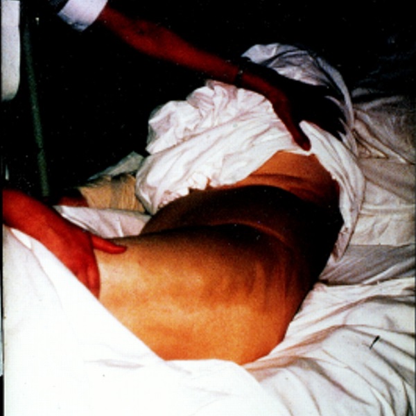
Use of this content is subject to our disclaimer