Images and videos
Images
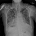

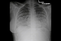
Acute heart failure
Chest x-ray showing acute pulmonary oedema with increased alveolar markings and bilateral pleural effusions
From the private collections of Syed W. Yusuf, MBBS, MRCPI, and Daniel Lenihan, MD
See this image in context in the following section/s:
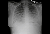
Acute heart failure
Chest x-ray showing acute pulmonary oedema with increased alveolar markings, fluid in the horizontal fissure, and blunting of the costophrenic angles
From the private collections of Syed W. Yusuf, MBBS, MRCPI, and Daniel Lenihan, MD
See this image in context in the following section/s:
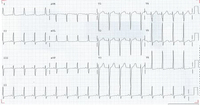
Acute heart failure
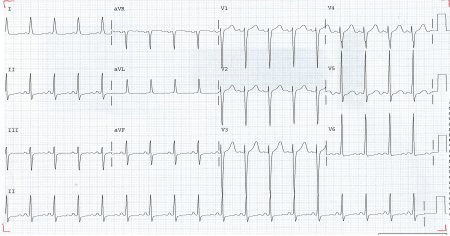
ECG showing left ventricular hypertrophy with sinus tachycardia
From the private collections of Syed W. Yusuf, MBBS, MRCPI, and Daniel Lenihan, MD; used with permission
See this image in context in the following section/s:
Acute heart failure
Chest x-ray showing acute pulmonary oedema with increased alveolar markings and bilateral pleural effusions
From the private collections of Syed W. Yusuf, MBBS, MRCPI, and Daniel Lenihan, MD; used with permission
See this image in context in the following section/s:
Acute heart failure
Chest x-ray showing acute pulmonary oedema with increased alveolar markings, fluid in the horizontal fissure, and blunting of the costophrenic angles
From the private collections of Syed W. Yusuf, MBBS, MRCPI, and Daniel Lenihan, MD; used with permission
See this image in context in the following section/s:
Acute heart failure
ECG showing left ventricular hypertrophy with sinus tachycardia
From the private collections of Syed W. Yusuf, MBBS, MRCPI, and Daniel Lenihan, MD
See this image in context in the following section/s:
Use of this content is subject to our disclaimer