Images and videos
Images

Assessment of dysuria
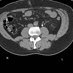
CT scan showing transitional cell carcinoma of the bladder blocking the right ureteric orifice
From the personal collection of Dr Kasra Saeb-Parsy
See this image in context in the following section/s:

Assessment of dysuria
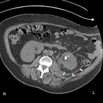
CT scan showing left ureteric calculi
From the personal collection of Dr Kasra Saeb-Parsy
See this image in context in the following section/s:

Assessment of dysuria
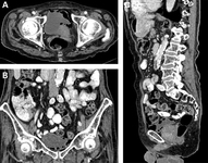
Emphysematous cystitis: (A) horizontal CT slice showing increased emphysema; (B) coronal CT slice showing increased emphysema; (C) sagittal CT slice showing increased emphysema
Middela S, Green E, Montague R. Emphysematous cystitis: radiological diagnosis of complicated urinary tract infection. BMJ Case Rep. 2009; doi:10.1136/bcr.05.2009.1832. Used with permission
See this image in context in the following section/s:

Assessment of dysuria
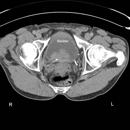
CT scan showing left renal stone (white arrow) with perinephric stranding around the left kidney (blue chevrons) and pyelonephritis
From the personal collection of Dr Kasra Saeb-Parsy
See this image in context in the following section/s:
Use of this content is subject to our disclaimer