Images and videos
Images

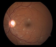


Hypertensive emergencies
Fundus photograph of the left eye with multiple cotton-wool spots typical of hypertensive retinopathy
Courtesy Angie Wen MD, Attending Faculty, New York Eye and Ear Infirmary, New York; used with permission
See this image in context in the following section/s:
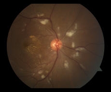
Hypertensive emergencies
Fundus photograph of the right eye centred on the optic nerve, showing multiple cotton-wool spots and macular exudates in a radiating star configuration around the fovea
Courtesy Angie Wen MD, Attending Faculty, New York Eye and Ear Infirmary, New York; used with permission
See this image in context in the following section/s:
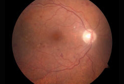
Hypertensive emergencies
Fundus photograph of the right eye with multiple dot-blot haemorrhages typical of hypertensive retinopathy
Courtesy Angie Wen MD, Attending Faculty, New York Eye and Ear Infirmary, New York; used with permission
See this image in context in the following section/s:
Videos
 Venepuncture and phlebotomy animated demonstration
How to take a venous blood sample from the antecubital fossa using a vacuum needle.
 How to perform an ECG animated demonstration
How to record an ECG. Demonstrates placement of chest and limb electrodes.
Use of this content is subject to our disclaimer