Images and videos
Images

Testicular cancer
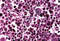
Non-seminoma
From the collection of Dr Francisco G. La Rosa; used with permission
See this image in context in the following section/s:

Testicular cancer
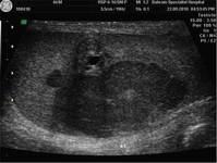
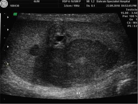
Longitudinal grey-scale image of the medial right testicle shows a 5.4×5.6 cm intratesticular heterogenous focus with a peripheral hypoechoic rim
BMJ Case Reports 2010; doi:10.1136/bcr.06.2010.3119
See this image in context in the following section/s:
Testicular cancer
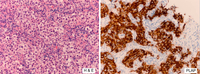
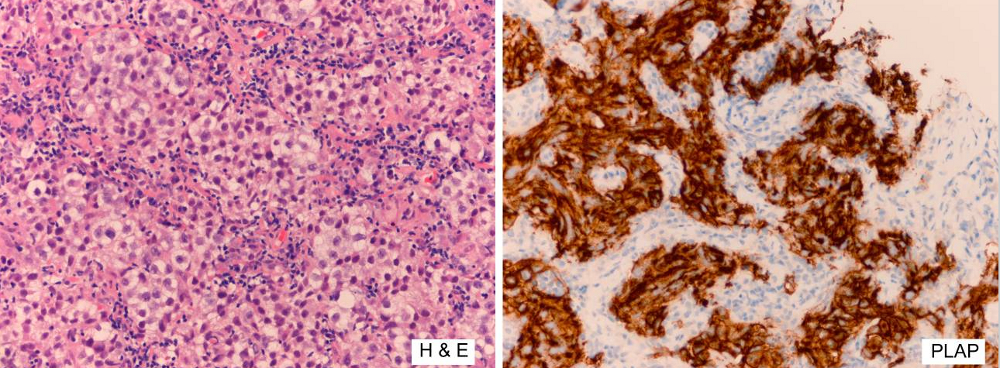
Metastatic seminoma. Left image: H&E stain low power. Right image placental alkaline phosphatase (PLAP) stain high power. The H&E image shows nests of malignant cells with clear cytoplasm; the tumour cells are strongly positive for PLAP confirming the diagnosis
Law C et al. BMJ Case Rep. 2020 Apr 22;13(4):e233368. doi: 10.1136/bcr-2019-233368; used with permission
See this image in context in the following section/s:
Testicular cancer
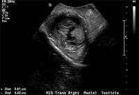
Ultrasonographic picture of testicular mass as hypoechoic lesion
BMJ Case Reports 2011; doi:10.1136/bcr.12.2010.3565
See this image in context in the following section/s:
Use of this content is subject to our disclaimer