Approach
The work-up should include:
Clinical symptoms compatible with Raynaud's phenomenon (RP)
A history to determine the frequency/severity of the attacks
Smoking history
Review of systems
Family history of RP and other connective tissue disease
Physical examination focusing on features of RP and associated connective tissue disease
Investigations may include FBC, erythrocyte sedimentation rate (ESR) or C-reactive protein, antinuclear antibody (ANA), and urinalysis
Referral to a rheumatologist or other specialist if necessary (i.e., for determining if RP is secondary to a connective tissue disease and/or help with respect to treatment in severe RP).
History
RP is diagnosed following a thorough history eliciting features of pallor of digits with subsequent cyanosis and/or then rubor. It is normal for digits to turn mottled or cyanotic in the cold; pallor should be present to make the diagnosis. The history should focus on:
The location (fingertips or tips of the toes are the most common)
A clear demarcation of colour change that usually is circumferential around the digit
Cyanosis and/or rubor on rewarming.
[Figure caption and citation for the preceding image starts]: Cyanotic Raynaud's phenomenon (RP) in a patient with mixed connective tissue diseaseFrom the personal collection of Dr Janet Pope; used with permission [Citation ends].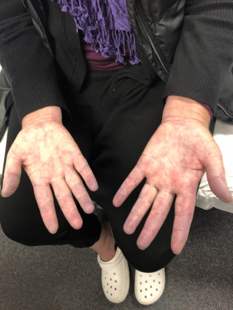 [Figure caption and citation for the preceding image starts]: Well-demarcated pallor of the 3rd finger in a patient with primary Raynaud’s phenomenon (RP)From the personal collection of Dr Janet Pope; used with permission [Citation ends].
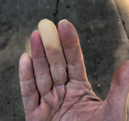
Indicators of increased severity of RP are attacks in warm weather (without exposure to cold or air conditioning), late onset RP (age >40 years), and the presence of complications such as digital ulcers.
A history of other systemic vasospasm symptoms such as migraine, irritable bowel, or anorexia may indicate primary RP, as well as increased vasodilatory responses to stimuli such as warmth, alcohol, and spicy food.[4]
A review of systems should rule out secondary causes such as connective tissue disease, which could include inflammatory arthritis, prolonged morning stiffness (such as in metacarpophalangeal, radiocarpal, and proximal interphalangeal joints), rash, photosensitivity, alopecia, significant dry eyes/dry mouth, puffiness of fingers or tightness of the skin, or significant oesophageal dysmotility/GORD.
A family history of RP and other connective tissue disease such as rheumatoid arthritis, systemic lupus erythematosus, scleroderma, Sjogren's syndrome, polymyositis, and juvenile idiopathic arthritis should be sought. Other causes of secondary RP may be obvious, such as malignancy, chemotherapeutic exposure, or severe peripheral vascular disease. Taking a clear drug and occupational history may also help diagnose secondary RP.[4]
RP can be confused with other vascular diseases, but biphasic or triphasic colour change is not present in these conditions. Onset of primary RP is rare after 40 years of age and often begins in adolescence. If the patient is young and has no signs/symptoms of connective tissue disease, primary RP should be suspected. In this instance, a good history and physical examination are often sufficient.
Examination
Physical examination should include:
An overview of general health and blood pressure (BP). Where asymmetrical RP is present, BP should be checked in both arms.[4]
Checking peripheral pulse to identify obstructive vascular disease. If RP sounds atypical (e.g., older age of onset of symptoms compatible with systemic disease), then blood pressure should be checked in both arms to rule out major discrepancies between arms, and the presence of carotid bruits should be checked. Giant cell arteritis and Takayasu's arteritis, for example, can be accompanied by large-vessel narrowing, which can affect blood pressure in a limb and can also produce carotid bruits.
Checking upper limb circulation to identify arterial occlusion. The Allen test should be performed by asking the patient to clench their fist and raise their arm, and pressure should be applied to the radial and ulnar arteries. Pressure should then be released one artery at a time, and colour should return to the hand when the patient opens their fingers. If pallor persists, this suggests occlusion of the uncompressed artery.[4] An abnormal test in a young patient is suggestive of Buerger's disease. See Buerger’s disease.
Checking for complications, including digital ulcers, digital pits (fibrotic plugs from ischaemia), digital tuft resorption, threatened ischaemic digit, gangrene of the fingertip/entire finger, infection including osteomyelitis, or auto-amputation. These are strong signs of secondary RP.[4]
A detailed skin examination to identify signs of an associated disorder (e.g., connective tissue disease), which can include telangiectasia, sclerodactyly, dactylitis or synovitis, malar rash, splinter haemorrhages, livedo reticularis, tightening of the skin in other areas (e.g., around the mouth), and alopecia areata.[4][6]
[Figure caption and citation for the preceding image starts]: Digital infarcts and nailfold changesFrom the personal collection of Dr Janet Pope; used with permission [Citation ends]. [Figure caption and citation for the preceding image starts]: Digital pits on the 3rd finger of the left handFrom the personal collection of Dr Janet Pope; used with permission [Citation ends].
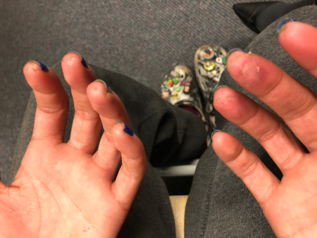 [Figure caption and citation for the preceding image starts]: Digital ulcer in a patient with secondary Raynaud’s phenomenon (RP)From the personal collection of Dr Janet Pope; used with permission [Citation ends].
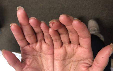 [Figure caption and citation for the preceding image starts]: Raynaud’s phenomenon (RP) and inflammatory arthritis with sclerodactyly distal to the proximal interphalangeal jointsFrom the personal collection of Dr Janet Pope; used with permission [Citation ends].
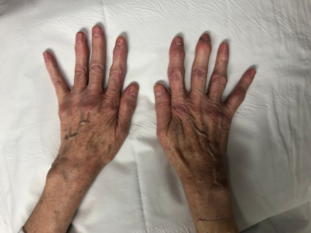 [Figure caption and citation for the preceding image starts]: Swollen/puffy fingers in a patient with secondary Raynaud’s phenomenon (RP)From the personal collection of Dr Janet Pope; used with permission [Citation ends].
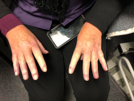 [Figure caption and citation for the preceding image starts]: Secondary Raynaud’s phenomenon (RP) with sclerodactylyFrom the personal collection of Dr Janet Pope; used with permission [Citation ends].
Early diagnosis and management of concomitant connective tissue disease is crucial in improving outcomes in RP.[4] The likelihood of underlying connective tissue disease increases when age of onset is more than 40 years.
Investigations
In most cases, a good history and physical examination are sufficient for diagnosis. However, as secondary RP may pre-date a connective tissue disease or may coincide with the onset of underlying conditions (e.g., diffuse cutaneous systemic sclerosis or polymyositis), all patients presenting with RP should undergo the following tests when available:[4]
ANA with titre and pattern
FBC
ESR or CRP
Urinalysis.
Capillary microscopy is usually not performed in primary care, but early referral to a rheumatologist who can perform this is recommended to exclude connective tissue disease.[4]
Follow-up should take place if a patient has:
Strongly positive ANA
Dilated capillaries at the nailbed
An uncommon ANA pattern such as an anti-centromere pattern
An elevated ESR and/or CRP (less predictive).
[Figure caption and citation for the preceding image starts]: Visible dilated capillaries at the nailbedFrom the personal collection of Dr Janet Pope; used with permission [Citation ends].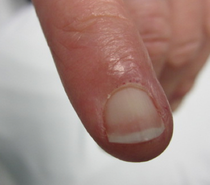
Follow-up is necessary in these patients as 30% will develop a connective tissue disease within the next 5 years. In particular, an anti-centromere antibody and/or dilated capillaries at the peri-ungual region are predictive for scleroderma.[28]
A referral to a rheumatologist should be considered if secondary RP is suspected, or if complications such as digital ulcers are present. The presence of digital ulceration is generally thought to exclude a diagnosis of primary RP, so a secondary cause should be sought if ulceration occurs.[4]
Rarely, RP can be caused by obstruction of blood flow by a cervical rib. This can be identified on chest x-ray, although this investigation is not carried out routinely on patients with mild RP and is ordered only if there is a high index of suspicion.
Use of this content is subject to our disclaimer