Images and videos
Images


Discogenic low back pain
Spondylolisthesis: flexion/extension views
From the collection of Dr N. Quiraishi
See this image in context in the following section/s:

Discogenic low back pain
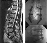
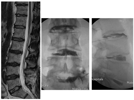
Pre- and post-surgical views: a patient presents with back pain and neurogenic claudication with stenosis and degenerative slip at L4-5 and a degenerate disc at L5S1 (left, T2-weighted sagittal MRI); L4-S1 decompression and instrumented fusion and a 2-level transforaminal lumbar interbody fusion was performed (AP radiograph top; lateral, bottom)
From the collection of Dr N. Quiraishi
See this image in context in the following section/s:
Discogenic low back pain
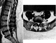
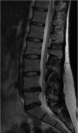
MRI spine: degenerate L4-5 disc with a disc bulge and L5S1 disc with a high-intensity zone
From the collection of Dr N. Quiraishi
See this image in context in the following section/s:
Discogenic low back pain
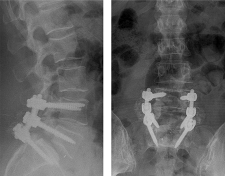
Postoperative radiographs
From the collection of Dr N. Quiraishi
See this image in context in the following section/s:
Discogenic low back pain
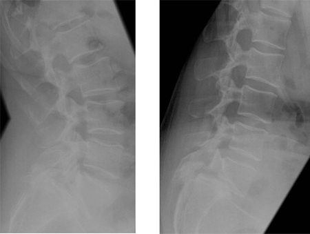
Discography: a patient presents with back and right leg pain that did not respond to a trial of conservative management. MRI (left, sagittal T2-weighted image) demonstrates multiple degenerate discs with loss of normal hydration, reduced signal, and loss of the nuclear-annular transition, with a normal disc height. A 3-level discography at L3-4, L4-5, and L5S1 (middle, AP radiograph; right, lateral radiograph) reveals a low-pressure injection with degenerate annular tears at all 3 levels, with 5/5 pain concordance at L5S1; 3/5 at L4-5; and 0/5 at L3-4
From the collection of Dr N. Quiraishi
See this image in context in the following section/s:

Discogenic low back pain
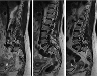
Preoperative MRI sagittal T2 sequence
From the collection of Dr N. Quiraishi
See this image in context in the following section/s:
Discogenic low back pain
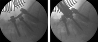
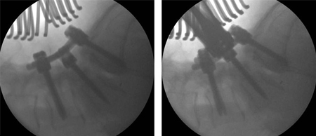
Intra-operative images showing a gradual reduction of the deformity: L4 to S1 instrumented fusion, transforaminal fusion at L5S1 and bilateral L5 decompression
From the collection of Dr N. Quiraishi
See this image in context in the following section/s:

Discogenic low back pain
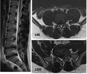
T2-weighted MRI spine: sagittal view (left) demonstrates degenerate discs; axial view (right) demonstrates left-sided L5S1 foraminal narrowing
From the collection of Dr N. Quiraishi
See this image in context in the following section/s:

Discogenic low back pain
MRI spine: degenerate facet joints and a large facet cyst from the left L5S1 facet joint compressing the left S1 nerve root
From the collection of Dr N. Quiraishi
See this image in context in the following section/s:

Discogenic low back pain
T2-weighted MRI spine: sagittal view (left) demonstrates 2 level disc dehydration at L4-5 and L5S1 with a moderate reduction in disc height; axial views (right) demonstrate constitutionally narrow canal at L4-5 with a moderate disc prolapse and a large disc prolapse at L5S1 level with left S1 root compression
From the collection of Dr N. Quiraishi
See this image in context in the following section/s:

Discogenic low back pain
Disc replacement: patient presents with severe back pain, having previously undergone right L5S1 discectomy for a right S1 radiculopathy. Though initially recovered, the right S1 pain recurred after 10 months, with back pain. An MRI scan shows a degenerate L5S1 disc (left, T2-weighted sagittal view). Patient subsequently had a disc replacement (AP radiograph top right, lateral bottom right). The pain in the back and the right leg resolved completely
From the collection of Dr N. Quiraishi
See this image in context in the following section/s:
Use of this content is subject to our disclaimer