Aetiology
The overall causative agents in IE are well documented and have been relatively stable, based on population-based studies over time. The most common pathogens are listed below; these together with any underlying risk factors indicate the most likely causative organism:[2][13]
Staphylococcus aureus (31% cases)
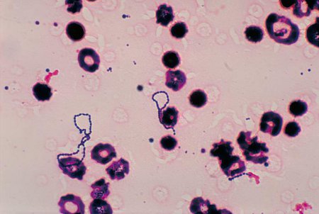
Viridans group streptococci [Figure caption and citation for the preceding image starts]: Photomicrograph of Streptococcus viridans bacteria that had been grown in a blood cultureCDC/Dr Mike Miller; used with permission [Citation ends].
Coagulase-negative staphylococci
Enterococci
Streptococcus gallolyticus (formerly Streptococcus bovis)
Oral streptococci (Streptococcus mitis, sanguinis, anginosus, salivarius, downei, and mutans)
Culture-negative Haemophilus species, Aggregatibacter, Cardiobacterium hominis, Eikenella corrodens, and Kingella species (HACEK)
Fungi
Haemophilus parainfluenzae
Coxiella burnetii
Brucella species.
Patients who develop native valve endocarditis in the absence of intravenous drug use commonly present with staphylococci, viridans group streptococci, or enterococci, with other pathogens being less frequent. Patients who use intravenous drugs often present with right-sided valvular involvement and are more likely to have S aureus, streptococci, gram-negative bacilli, or polymicrobial infections.[2]
Prosthetic valve endocarditis is most commonly caused by coagulase-negative staphylococci, S aureus, enterococci, or gram-negative bacilli. It should be noted that early prosthetic valve endocarditis is often caused by Staphylococcus epidermidis.[2]
Pathophysiology
IE typically develops on the valvular surfaces of the heart, which have sustained endothelial damage secondary to turbulent blood flow. As a result, platelets and fibrin adhere to the underlying collagen surface and create a prothrombotic milieu. Bacteraemia leads to colonisation of the thrombus and perpetuates further fibrin deposition and platelet aggregation, which develops into a mature infected vegetation.[14]
Acute IE is usually associated with more virulent organisms, classically Staphylococcus aureus. Thrombus is formed by the offending organism and S aureus may invade endothelial cells and increase the expression of adhesion molecules as well as prothrombotic factors.[15]
Classification
Clinical classification[1]
Acute symptoms typically develop over a period of days and for up to 6 weeks; include spiking fevers, tachycardia, fatigue, and progressive damage to cardiac structures.
Subacute: symptoms occur over the course of weeks to months. Patients will often describe vague constitutional symptoms.
Chronic: symptoms continue for more than 3 months.
Classification by nidus or location of infection
Native valve endocarditis (NVE)
Patients who develop NVE in the absence of intravenous drug use commonly present with viridans group streptococci, enterococci, or staphylococci, with other pathogens being less frequent. Patients who use intravenous drugs often present with right-sided valvular involvement and are more likely to have Staphylococcus aureus, streptococci, gram-negative bacilli, or polymicrobial infections.[2]
Prosthetic valve endocarditis (PVE)
Between 10% and 30% of all patients with IE have prosthetic heart valves.[2][3] PVE is defined as being early or late depending on whether it arises within 1 year after valve replacement (early) or after 1 year (late). The focus is placed on the likely causative organisms involved at different time points in the disease process. Early PVE is often caused by S aureus or coagulase-negative staphylococci.[4] Late PVE tends to be caused by the same organisms as NVE, and non-surgical treatment in this group may be effective.
Device-related endocarditis
The increasingly widespread use of implanted devices has contributed to the rising incidence of this complication, though it remains relatively rare overall. Estimates of incidence vary but it is a serious complication that is often difficult to diagnose and carries with it significant mortality.[5][6]
Although clinically challenging, distinction should be made between local device infection and device-related IE.
Risk factors for device-related IE include renal failure, haematoma at the site of implantation, diabetes mellitus, and anticoagulation.
Right-sided endocarditis
Infections of right-sided heart valves make up 5% to 10% of the total number of patients with IE, and are most commonly associated with patients who use intravenous drugs.[7][8] S aureus is the most common causative organism and accounts for 60% to 90% of patients with IE.[9][10] The tricuspid valve is the most commonly affected, although the pulmonary valve is also susceptible to infection.[8]
Use of this content is subject to our disclaimer