Images and videos
Images
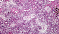


Organising pneumonia
Medium-powered pathology slide showing circular and branching bronchioles filled with polypoid plugs of granulation tissue and alveoli filled with organising pneumonia
From the collection of Gary R. Epler, MD
See this image in context in the following section/s:
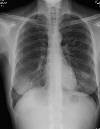
Organising pneumonia
Chest x-ray showing bilateral patchy infiltrates
From the collection of Gary R. Epler, MD
See this image in context in the following section/s:
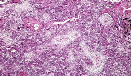
Organising pneumonia
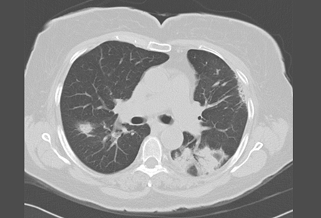
High-resolution chest CT showing bilateral ground glass opacities and a posterior triangular-based infiltrate with an air bronchogram
From the collection of Gary R. Epler, MD
See this image in context in the following section/s:
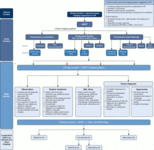
Organising pneumonia
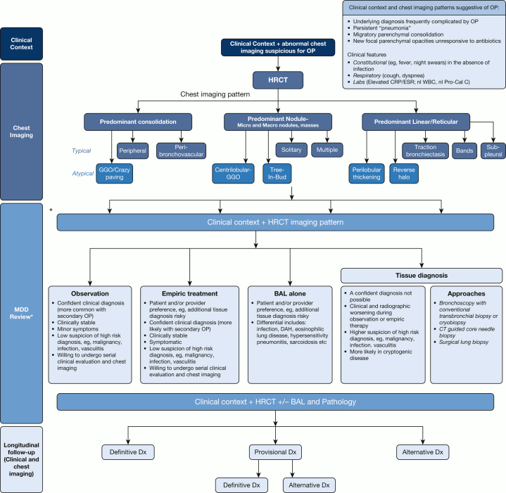
Algorithmic approach to organizing pneumonia. ∗ A formal MDD may not be required in all cases, especially if the combination of clinical context and radiographic pattern is sufficiently convincing of the OP diagnosis. In such cases, a discussion between the physician and the radiologist is strongly encouraged. CRP = C-reactive protein; DAH = diffuse alveolar hemorrhage; Dx = diagnosis; ESR = erythrocyte sedimentation rate; GGO = ground-glass opacification; HRCT = high-resolution CT; MDD = multidisciplinary discussion; nl Pro-Cal C = normal procalcitonin; nl WBC = normal WBC; OP = organizing pneumonia
Cherian SV, et al. Chest. 2022 Jul;162(1):156-78. doi: 10.1016/j.chest.2021.12.659. Epub 2022 Jan 14; used with permission
See this image in context in the following section/s:
Videos
 Late inspiratory crackles (rales)
Auscultation sounds: late inspiratory crackles (rales)
Use of this content is subject to our disclaimer