Images and videos
Images
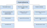
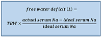
Hypernatraemia
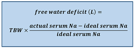
Free water deficit formula. TBW (total body water) = patient body weight (kg) x 0.5 (women/older men) or 0.6 (young men or children) or 0.4 (dehydrated patients). Na = sodium
Created by the BMJ Knowledge Centre
See this image in context in the following section/s:
Hypernatraemia
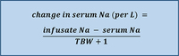
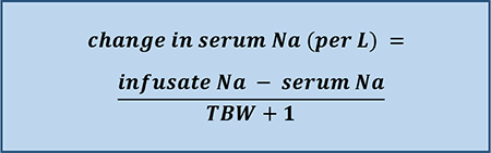
Adrogué-Madias formula. TBW (total body water) = patient body weight (kg) x 0.5 (women/older men) or 0.6 (young men or children) or 0.4 (dehydrated patients). Na = sodium. Sodium concentration of common fluids (per litre): normal saline (0.9%) - 154 mmol/L; lactated Ringer's solution - 130 mmol/L; half-normal saline (0.45%) - 77 mmol/L; dextrose 5% in water - 0 mmol/L; enteral water - 0 mmol/L
Created by the BMJ Knowledge Centre
See this image in context in the following section/s:
Hypernatraemia
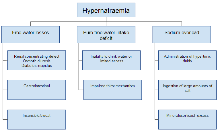
Possible aetiologies of hypernatraemia
Created by the BMJ Knowledge Centre
See this image in context in the following section/s:
Hypernatraemia
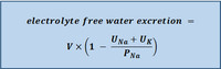
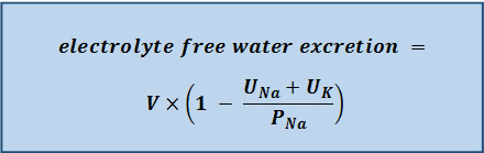
Electrolyte-free water excretion formula. V = urine flow rate. UNa = urine concentration of sodium (mmol/L). UK = urine concentration of potassium (mmol/L). PNa = plasma concentration of sodium (mmol/L)
Created by the BMJ Knowledge Centre
See this image in context in the following section/s:

Hypernatraemia
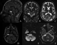
Brain MRI showing a symmetric central pontine lesion (arrows), sparing the peripheral fibres, with a typical trident shape and areas of restricted diffusion. Suggests osmotic demyelination syndrome. Note the symmetric thalamic lesions (open arrows) in images B and C
BMJ Case Reports 2012, doi: 10.1136/bcr:11.2011.5198
See this image in context in the following section/s:
Hypernatraemia
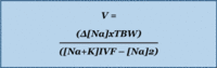
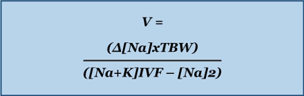
V = volume needed. Na = sodium. TBW (total body water) = patient body weight (kg) x 0.5 (women/older men) or 0.6 (young men or children) or 0.4 (dehydrated patients). K = potassium. [Na]2 = the desired change in sodium concentration
Created by the BMJ Knowledge Centre
See this image in context in the following section/s:

Hypernatraemia
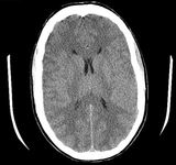
CT brain showing diffuse sulcal effacement and slight blurring of grey-white differentiation consistent with global cerebral oedema. There is no evidence of herniation
Dr Hari Trivedi, Department of Radiology, San Francisco General Hospital, University of California; used with permission.
See this image in context in the following section/s:
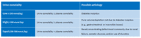
Hypernatraemia
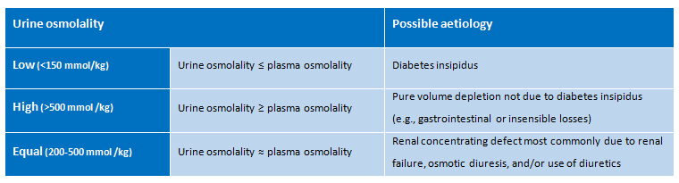
Possible aetiologies of hypernatraemia based on urine osmolality
Created by the BMJ Knowledge Centre
See this image in context in the following section/s:
Use of this content is subject to our disclaimer