Images and videos
Images


Carotid artery stenosis
MRI of carotid artery. The arrow shows narrowing of the lumen of the carotid artery caused by intramural haematoma
Used with permission from BMJ Case Reports 2012; doi:10.1136/bcr.01.2012.5636
See this image in context in the following section/s:

Carotid artery stenosis
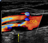
Echolucent internal carotid artery atheroma (yellow arrow) causing 70% stenosis (North American Symptomatic Carotid Endarterectomy Trial criteria). CCA = common carotid artery, ECA = external carotid artery, ICA = internal carotid artery, STA = superior thyroid artery
Used with permission from BMJ 2013;346:f2420
See this image in context in the following section/s:

Carotid artery stenosis
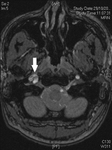
Magnetic resonance time of flight image of brain. The arrow shows the right carotid artery with a crescent-shaped appearance. This is consistent with intramural haematoma consequent upon dissection of the right carotid artery
Used with permission from BMJ Case Reports 2012; doi:10.1136/bcr.01.2012.5636
See this image in context in the following section/s:

Carotid artery stenosis
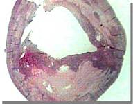
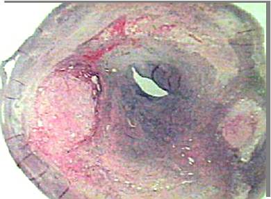
Ruptured plaque with intraplaque haemorrhage explanted from a patient with carotid stenosis and a recent atheroembolic stroke
From the personal collection of Brajesh K. Lal, MD
See this image in context in the following section/s:
Carotid artery stenosis
Stable plaque with a small deep-seated lipid core explanted from a patient with a high-grade carotid stenosis without neurological symptoms
From the personal collection of Brajesh K. Lal, MD
See this image in context in the following section/s:
Videos
 Carotid bruit
Auscultation: Carotid bruit
Use of this content is subject to our disclaimer