Images and videos
Images

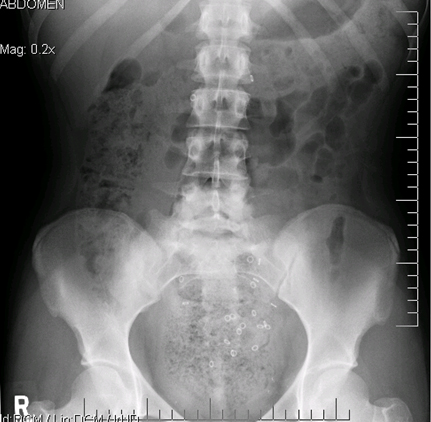
Assessment of nausea and vomiting in children
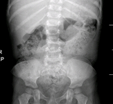
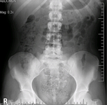
Abdominal x-ray showing faecal impaction in a patient with constipation
From the collections of Dr R.A. Gomez-Suarez and Dr J.E. Fortunato; used with permission
See this image in context in the following section/s:
Assessment of nausea and vomiting in children
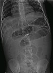
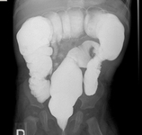
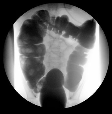
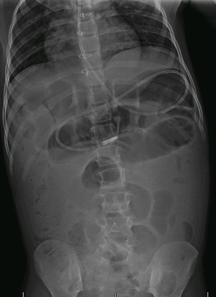
Abdominal x-ray showing filled right colon and empty left colon and rectum in a patient with intussusception
From the collections of Dr R.A. Gomez-Suarez and Dr J.E. Fortunato; used with permission
See this image in context in the following section/s:

Assessment of nausea and vomiting in children
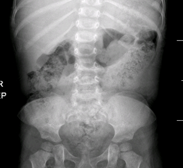
Abdominal x-ray showing intestinal malrotation; note the small bowel is located to the right side of the midline
From the collections of Dr R.A. Gomez-Suarez and Dr J.E. Fortunato; used with permission
See this image in context in the following section/s:
Assessment of nausea and vomiting in children
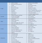
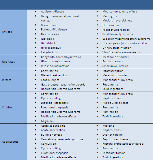
Aetiology of nausea and vomiting in children and adolescents grouped according to age
From the collections of Dr R.A. Gomez-Suarez and Dr J.E. Fortunato; used with permission
See this image in context in the following section/s:

Assessment of nausea and vomiting in children
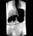
Barium enema showing transition zones in patients with Hirschsprung's disease
From the collections of Dr R.A. Gomez-Suarez and Dr J.E. Fortunato; used with permission
See this image in context in the following section/s:
Assessment of nausea and vomiting in children
Abdominal x-ray showing small bowel volvulus, a common cause of bilious vomiting
From the collections of Dr R.A. Gomez-Suarez and Dr J.E. Fortunato; used with permission
See this image in context in the following section/s:
Assessment of nausea and vomiting in children
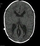
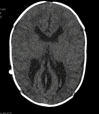
CT showing increased volume of lateral ventricles secondary to non-communicating hydrocephalus
From the collections of Dr R.A. Gomez-Suarez and Dr J.E. Fortunato; used with permission
See this image in context in the following section/s:
Assessment of nausea and vomiting in children
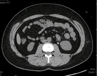
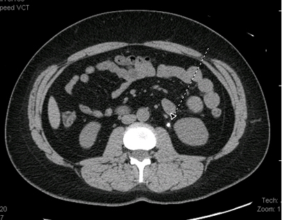
CT abdomen showing small calcification in area of the left ureteral space, which corresponds to the presence of a kidney stone
From the collections of Dr R.A. Gomez-Suarez and Dr J.E. Fortunato; used with permission
See this image in context in the following section/s:
Assessment of nausea and vomiting in children
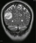
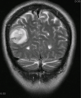
CT head showing right parietotemporal brain tumour
From the collections of Dr R.A. Gomez-Suarez and Dr J.E. Fortunato; used with permission
See this image in context in the following section/s:
Assessment of nausea and vomiting in children
Sitz marker test showing retention of ingested markers in the rectosigmoid region in a patient with constipation
From the collections of Dr R.A. Gomez-Suarez and Dr J.E. Fortunato; used with permission
See this image in context in the following section/s:
Use of this content is subject to our disclaimer