Approach
Dengue fever is a notifiable condition. In dengue-endemic regions, suspected, probable, and confirmed cases of dengue infection should be reported to the relevant authorities as soon as possible so that appropriate measures can be instituted to prevent dengue transmission.[2]
Early diagnosis is difficult as the clinical picture is similar to many other viral and bacterial infections. Re-emerging arboviral infections such as chikungunya and Zika virus are clinically indistinguishable from dengue without laboratory testing, and it is important to differentiate between these infections as they can produce similar symptoms, particularly during the acute phase.
Patients with dengue may be asymptomatic or present with undifferentiated fever (viral syndrome). Severity of infection can range from mild illness to severe disease. When a patient presents with either dengue haemorrhagic fever (DHF) or dengue shock syndrome (DSS), it may be difficult to differentiate these from other causes of shock. Therefore, laboratory studies play a valuable role in diagnosis from the first day of infection. A diagnosis of DF should be considered in any patient presenting with fever, generalised skin flushing, leukopenia, and thrombocytopenia.
A high index of clinical suspicion is required as early clinical findings may be non-specific. Arriving at a correct diagnosis early in the course of the infection is important for management and preventing complications.
History
A diagnosis of DF should be suspected in any patients residing in countries where dengue infection is endemic, or travelling from such areas within the past 2 weeks.
Approximately 75% of infections are asymptomatic. Symptomatic infection most commonly presents as a mild to moderate non-specific acute febrile illness. Approximately 5% of patients progress to severe, life-threatening disease.[73]
After the incubation period (4 to 10 days), the onset of symptoms is usually abrupt in those with symptomatic infection. Fever is characteristic; very often it has an abrupt onset with very high spikes of 39.4°C to 40.5°C (103°F to 105°F). It may also be biphasic and have a remittent pattern or be low grade. The fever generally lasts approximately 5 to 7 days, and may cause febrile seizures or delirium in young children. Rapid defervescence may indicate that a patient with dengue infection is about to enter the critical phase of infection.[2][16]
Aches and pains, particularly backache, arthralgia, myalgia, and bone pain, are common. Headache is also common and is usually constant and towards the front of the head. It usually improves within a few days. Severe retro-orbital pain on eye movement or with little pressure applied to the eyeball is also common. The most commonly reported symptoms are fever (98%), headache (76%), malaise (76%), and asthenia (74%).[18]
Gastrointestinal symptoms (e.g., anorexia, nausea/vomiting, epigastric discomfort/pain), lethargy/restlessness, collapse, or dizziness may also be present. Often the patient will report a lack of appetite and/or changes to taste sensation. Gastrointestinal symptoms, weakness, and dizziness may be more marked in DHF.
Upper respiratory tract symptoms (e.g., cough, sore throat) are usually absent; however, may atypically occur in mild infection.
Physical examination
Diffuse skin flushing of the face, neck, and chest develop early on in the infection. This evolves into a maculopapular or rubelliform rash involving the whole body, usually on the third or fourth day of the fever. The flushing may blanch when the affected skin is pressed.[74] The rash fades with time and appears as islands of pallid areas during the convalescent phase. The flushing/rash may not be seen, may be difficult to see, or may look different in people with black skin.[Figure caption and citation for the preceding image starts]: Typical skin flushing with islands of normal skin in patient with dengue feverFrom the collection of Professor S.A.M. Kularatne [Citation ends].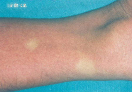
Haemorrhagic signs include petechiae, purpura, or a positive tourniquet test (performed by inflating a blood pressure cuff to a point midway between systolic and diastolic blood pressures for 5 minutes; the test is positive if ≥10 petechiae per square inch appear on the forearm). More significant haemorrhage can manifest as epistaxis, gingival bleeding, haematemesis, melaena, vaginal bleeding (in women of childbearing age), or bleeding from a venipuncture site. These signs can occur with either DF or DHF.[1][2][Figure caption and citation for the preceding image starts]: Positive tourniquet test showing presence of rubelliform rash and petechiaeFrom the collection of Professor S.A.M. Kularatne [Citation ends]. [Figure caption and citation for the preceding image starts]: Autopsy specimen showing haemorrhage of the lungFrom the collection of Professor S.A.M. Kularatne [Citation ends].
 [Figure caption and citation for the preceding image starts]: Autopsy specimen showing subcutaneous bleedingFrom the collection of Professor S.A.M. Kularatne [Citation ends].
Hepatomegaly may be present. Plasma leakage is a sign of DHF, and clinical evidence of this includes the presence of ascites, postural dizziness, or pleural effusion.[1][2]
Circulatory collapse (i.e., cold clammy skin, rapid and weak pulse with narrowing of pulse pressure <20 mmHg with decreased diastolic pressure, postural drop of blood pressure >20 mmHg, capillary refill time >3 seconds, reduced urine output) indicates the presence of shock and supports a diagnosis of DSS.[1][2]
Unusual presentations and complications are uncommon, and the clinical picture will depend on the organ system affected.[1] Rarely, dengue infections may be accompanied by concurrent bacteraemia. Risk factors may include severe dengue infection, older age, and the presence of comorbidities.[4]
Phases of infection
Dengue infection has 3 distinct phases:[2]
Febrile
Critical
Convalescent.
The febrile phase is characterised by a sudden high-grade fever and dehydration that can last from 2 to 7 days and may be biphasic. Other signs and symptoms during this phase may include headache, retro-orbital eye pain, aches and pains, rash, and minor haemorrhagic manifestations.[2][73]
The critical phase is characterised by plasma leakage, bleeding, shock, and organ impairment and lasts for approximately 24 to 48 hours. It usually starts late in the febrile phase around the time of defervescence (although this does not always occur), around days 3 to 7 of the infection. The following warning signs indicate that a patient with dengue infection is about to enter the critical phase of infection:[2][16]
Abdominal pain or tenderness
Persistent vomiting
Clinical fluid accumulation (e.g., ascites, pleural effusion)
Mucosal bleeding
Lethargy/restlessness
Liver enlargement >2 cm
Laboratory: increase in haematocrit with rapid decrease in platelet count.
Most patients improve during the critical phase. However, those with substantial plasma leakage can develop severe disease within a few hours, with pleural effusions, ascites, hypoproteinemia, or haemoconcentration. Patients may appear to be well despite early signs of shock, but once hypotension develops, blood pressure declines rapidly. Patients may develop severe haemorrhagic manifestations during this phase.[73]
The convalescent phase is characterised by plasma leakage subsiding as the patient begins to reabsorb extravasated intravenous fluids and pleural and abdominal effusions. Haemodynamic status begins to stabilise, diuresis ensues, and the patient’s well-being improves. The rash may desquamate and become pruritic.[73]
Patients with DHF/DSS go through all 3 stages; however, patients with DF bypass the critical phase.[1][2][75]
Laboratory investigations
Laboratory tests should be ordered in all patients presenting with signs and symptoms that suggest infection with the dengue virus. The specific tests ordered will depend on which tests are available in the local area. In resource-poor regions, confirmatory laboratory tests are usually not available, and basic laboratory tests should be ordered (i.e., FBC including haematocrit, and liver function tests [LFTs]).
The results of these tests should be interpreted with caution, taking the clinical picture into account. For example, if a patient presents with an acute febrile illness and skin flushing and is from an area where dengue infection is endemic, the probability of dengue infection is high, and the results of basic laboratory tests may be used to support the diagnosis. However, confirmatory laboratory tests should always be ordered when possible, to rule out any differential diagnoses.
Initial laboratory investigations
An FBC should be ordered initially in all symptomatic patients. Typically, leukopenia and thrombocytopenia occur as early as the second day of fever.[1] Leukopenia, in combination with a positive tourniquet test, in a dengue-endemic area has a positive predictive value of 70% to 80%.[76][77] Leukopenia (with neutropenia) persists throughout the febrile period. In classical DF, thrombocytopenia is usually mild, although it may also be severe.[1]
Haematocrit may also rise approximately 10% in patients with DF due to dehydration.[1] LFTs are usually elevated, particularly alanine and aspartate aminotransferases.[1] Clotting studies are not required for diagnosis, but may play a useful role in the management of the infection in patients with haemorrhagic signs.
Laboratory criteria for diagnosis of DSS/DHF include:[1][2]
Rapidly developing, severe thrombocytopenia
Decreased total WCC and neutrophils and changing neutrophil-to-lymphocyte ratio
Elevated haematocrit (i.e., 20% increase from baseline is objective evidence of plasma leakage)
Hypoalbuminaemia (i.e., serum albumin <35 g/L [3.5 g/dL] suggests plasma leakage)
Elevated LFTs (i.e., aspartate aminotransferase [AST]:alanine aminotransferase [ALT] >2).
Monitoring platelet count, serum albumin, and AST and ALT levels during the febrile phase of the illness may improve early prediction of progression to severe dengue.[51] Thrombocytopenia and elevated AST level in the first 72 hours of fever onset are independent markers for predicting the development of severe dengue, based on moderate-certainty evidence.[78]
Confirmatory laboratory investigations
Confirmatory tests should always be ordered if available. This is important because DF can be confused with many non-dengue illnesses. The testing recommendations below are based on guidance from the World Health Organization.[1][2]
There are 4 types of diagnostic test available for confirmation of dengue virus infection:
Virus isolation
Viral antigen detection
Viral nucleic acid detection
Serology (antibody response).
The choice of test depends on numerous factors, including local availability, cost, time of sample collection, available facilities, and technical expertise. While direct methods such as viral nucleic acid or viral antigen detection are more specific, they are more costly and labour-intensive. Indirect methods (i.e., serology) are less specific, but are more accessible, faster, and less costly.[2] The identification of viral nucleic acid or viral antigen, plus the detection of an antibody response (serology), is preferable to either approach alone, if this is possible.[2] Viral nucleic acid or viral antigen detection is primarily used in the first 5 days of illness, and serological tests after the fifth day. Some tests differentiate between viral serotypes; however, this is not useful clinically.
Virus isolation is possible during the initial viraemic phase; however, this is only available in some locations and results are usually not available in a clinically meaningful time frame, so this test is generally not recommended. Reverse transcriptase-polymerase chain reaction (RT-PCR), serology (single serum specimen), and NS1 antigen test accurately identified over 90% of primary and secondary dengue cases during the first 10 days of illness in one study.[79]
Viral antigen detection:
Detection of the non-structural protein 1 (NS1) using enzyme-linked immunosorbent assay (ELISA) or rapid kits is useful in early diagnosis and can be ordered from days 1 to 5 of illness.[80] A serum specimen should be used. A positive result confirms diagnosis.
Advantages: easy to perform; rapid tests can be used in the field and provide results in a few hours; early diagnosis is possible, which may impact management.[2]
Disadvantages: may be as sensitive as viral nucleic acid detection; however, does not identify serotype.[2]
Viral nucleic acid detection:
RT-PCR is the method of choice and can be ordered in the first 5 to 7 days of fever onset. Tissue, whole blood, serum, plasma, or cerebrospinal fluid specimens can be used. A positive result confirms diagnosis.[81]
Advantages: the most sensitive and specific test available, especially in early infection; early diagnosis is possible, which may impact management; can identify serotype.[2]
Disadvantages: expensive, requires laboratory facilities and expertise, not rapid (takes 24 to 48 hours), cannot differentiate between primary and secondary infection, potential for false-positive result due to contamination.[2]
Serology:
Serology may be negative in the first 5 days of illness; therefore, IgM ELISA and IgG ELISA are the tests of choice after the first 5 days of illness (PCR is more sensitive in the first 5 days). The presence of IgG in the first few days of infection strongly suggests a secondary infection. Positive IgM and IgG in a single serum sample is highly suggestive of dengue infection, while IgM or IgG seroconversion in paired sera or a fourfold IgG titre increase in paired sera confirms the diagnosis.[2] Whole blood, serum, or plasma specimen can be used.
IgM rapid tests are commercially available and easy to use; however, their accuracy is poor as cross-reaction with other infectious agents and in autoimmune disorders can occur.
Haemagglutination-inhibition (HI) test is useful for diagnosing secondary dengue infection (i.e., titre ≥1:1280).
Advantages: inexpensive, easy to perform, more readily available in dengue-endemic areas, can distinguish between primary and secondary infection (i.e., IgM:IgG ratio <1.2 suggests secondary infection).[2]
Disadvantages: lower specificity compared to other tests, requires 2 serum samples, delays confirmation of diagnosis.[2]
The Centers for Disease Control and Prevention (CDC) recommends testing in anyone who is symptomatic and lives in, or has travelled to, areas where dengue virus is transmitted. They do not recommend testing in asymptomatic people or for preconception screening.[82]
An RT-PCR or NS1 test plus a serologic test is recommended during the first 7 days of illness (acute phase). If the RT-PCR or NS1 result is positive, the diagnosis is confirmed. If the RT-PCR or NS1 result is negative and the serology is positive, the diagnosis is presumptive.
Serology is recommended more than 7 days after symptom onset. If the RT-PCR or NS1 result and the serology from the acute phase samples were negative, a convalescent serum sample should be tested for antibodies. If the serology result is positive, the diagnosis is presumptive.
If the patient is pregnant and symptomatic, test for Zika virus infection in addition to dengue. Zika virus infection is known to be associated with microcephaly in newborn babies.[83] See Zika virus infection.
Testing recommendations vary between countries. Consult your local guidance for more information.
Imaging
Imaging studies are only required if DHF/DSS is suspected. A lateral decubitus chest X-ray of the right-hand side of the chest can be ordered to detect clinically undetectable pleural effusion in the early phase of plasma leakage. Ultrasound of the abdomen is very useful to detect the presence of ascites and plasma leak or other pathological changes in abdominal organs including the liver, gallbladder (i.e., oedema may precede plasma leakage), and kidneys.[1][2]
Emerging investigations
A newly developed pan-dengue virus reverse transcription-insulated isothermal PCR assay (RT-iiPCR) in combination with a nucleic acid analyser, may provide a highly reliable, sensitive, and specific point-of-need diagnostic assay in the future. Go YY, Rajapakse RP, Kularatne SA, et al. A pan-dengue virus reverse transcription-insulated isothermal polymerase chain reaction (RT-iiPCR) assay intended for point-of-need diagnosis of dengue infection using POCKITTM Nucleic Acid Analyzer.[84]
Differential diagnosis
It is important to differentiate between dengue, chikungunya, and Zika virus infection as the 3 diseases can produce similar symptoms, particularly during the acute phase. Co-infection with chikungunya (or both chikungunya and Zika) is also possible.[35]
The World Health Organization has produced a tool to help physicians differentiate between these 3 diseases.[85]
The Food and Drug Administration (FDA) has issued an Emergency Use Authorization for the Trioplex real-time RT-PCR assay. The assay allows physicians to tell if an individual is infected with dengue, Zika, or chikungunya virus using one test, instead of having to perform 3 separate tests.[86] Availability of this test varies depending on location.
Malaria infection (or co-infection) should also be considered. Jaundice (in dengue patients) and spontaneous bleeding (in malaria patients) should raise the suspicion of co-infection.[36]
See Differential Diagnosis section for more detailed information on how to differentiate between these infections.
Risk prediction tools
Algorithms for predicting the severity of dengue have been proposed; however, none have been adopted in clinical practice owing to differences in patient populations and variation across clinical definitions of dengue severity. Until these tools can be externally validated, they are not recommended in clinical practice.[87]

How to take a venous blood sample from the antecubital fossa using a vacuum needle.
Use of this content is subject to our disclaimer