Images and videos
Images


Assessment of breast mass
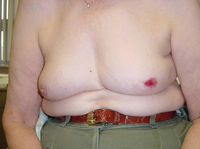
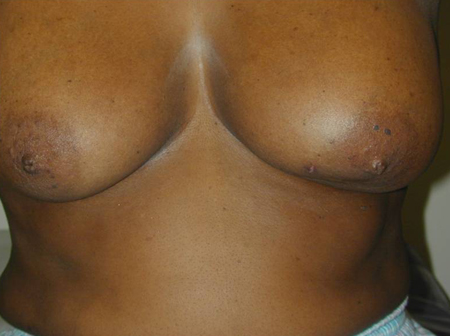
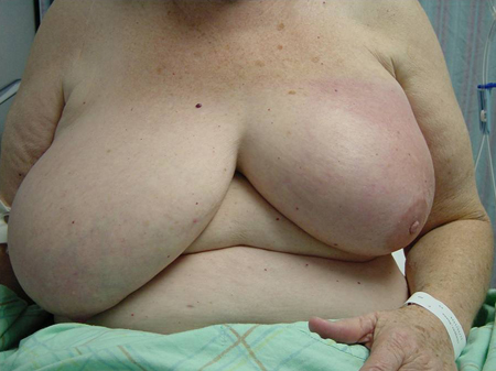
Patient with large breast mass and retraction at 6 o'clock of left breast, noted on elevating arms
Courtesy of Dr Anees Chagpar
See this image in context in the following section/s:
Assessment of breast mass
Excoriation of the nipple in a patient with Paget's disease
Courtesy of Dr Anees Chagpar
See this image in context in the following section/s:

Assessment of breast mass
Breast Imaging Reporting and Data System (BIRADS) criteria
Courtesy of Dr Anees Chagpar
See this image in context in the following section/s:

Assessment of breast mass
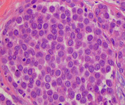
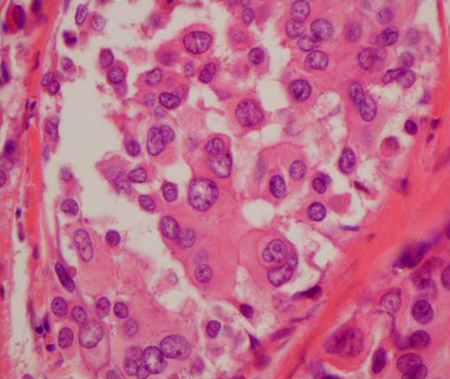
Histopathology of classic lobular carcinoma in situ (LCIS)
Courtesy of Dr Sunati Sahoo, University of Louisville; used with permission
See this image in context in the following section/s:
Assessment of breast mass
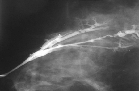
Ductogram demonstrating multiple intraductal papillomas
Courtesy of Dr Nancy Pile, University of Louisville; used with permission
See this image in context in the following section/s:
Assessment of breast mass
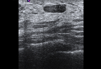
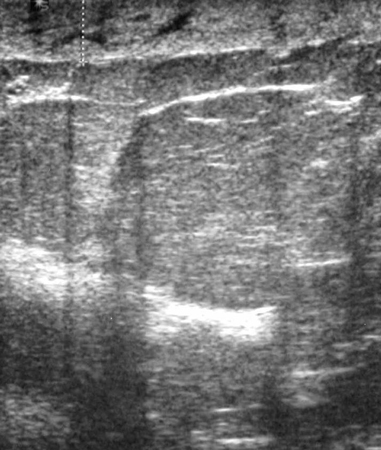
Ultrasonographic image of skin thickening in patient with inflammatory breast cancer
Courtesy of Dr Nancy Pile, University of Louisville; used with permission
See this image in context in the following section/s:
Assessment of breast mass
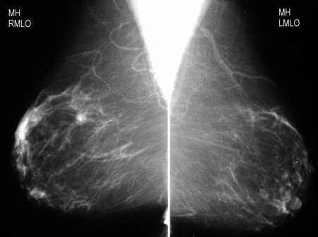
Screening mammogram demonstrating breast mass
Courtesy of Dr Nancy Pile, University of Louisville; used with permission
See this image in context in the following section/s:

Assessment of breast mass
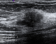
Ultrasonographic image of an invasive carcinoma
Courtesy of Dr Lane Roland, University of Louisville; used with permission
See this image in context in the following section/s:
Assessment of breast mass
Ultrasonographic image of a fibroadenoma
Courtesy of Dr Lane Roland, University of Louisville; used with permission
See this image in context in the following section/s:

Assessment of breast mass
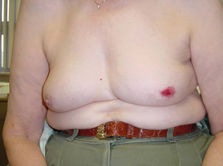
Obvious mass with skin involvement on left breast
Courtesy of Dr Anees Chagpar
See this image in context in the following section/s:

Assessment of breast mass
Magnification view demonstrating irregular spiculated mass with associated calcifications
Courtesy of Dr Nancy Pile, University of Louisville; used with permission
See this image in context in the following section/s:
Assessment of breast mass
Patient with inflammatory breast cancer who presented with a shrinking breast
Courtesy of Dr Anees Chagpar
See this image in context in the following section/s:
Assessment of breast mass
Histopathology of pleomorphic lobular carcinoma in situ (LCIS)
Courtesy of Dr Sunati Sahoo, University of Louisville; used with permission
See this image in context in the following section/s:

Assessment of breast mass
Breast biopsy techniques (FNA; fine needle aspiration)
Courtesy of Dr Anees Chagpar
See this image in context in the following section/s:
Assessment of breast mass
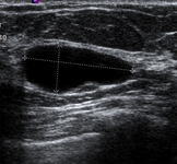
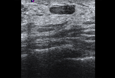
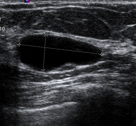
Ultrasonographic image of a simple cyst
Courtesy of Dr Lane Roland, University of Louisville; used with permission
See this image in context in the following section/s:

Assessment of breast mass
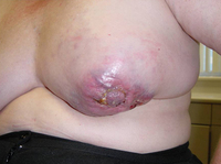
Obvious mass with skin involvement on right breast
Courtesy of Dr Anees Chagpar
See this image in context in the following section/s:

Assessment of breast mass
Ultrasonographic image of a complex cyst
Courtesy of Dr Lane Roland, University of Louisville; used with permission
See this image in context in the following section/s:
Assessment of breast mass
Diagnostic algorithm for breast ultrasound
Courtesy of Dr Anees Chagpar
See this image in context in the following section/s:
Use of this content is subject to our disclaimer