Aetiology
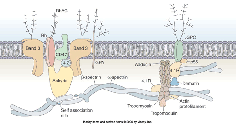
HS is an inherited disorder resulting in defects in the skeletal proteins of the red-cell membrane. These proteins are normally responsible for maintaining the red-cell shape and deformability.[14] Defects may occur in any of the 5 different membrane skeleton proteins:
Alpha-spectrin
Beta-spectrin
Ankyrin
Band 3 protein
Protein 4.2.
There are no other causative factors. [Figure caption and citation for the preceding image starts]: A schematic model of the organisation of the red-cell membrane (the proteins and lipids are not drawn to scale)This figure was published in: Young NS, Gerson SL, High KA, eds. Clinical haematology. Mohandas N, Reid ME, Erythrocyte structure, pp 36-38. Copyright Mosby Elsevier; 2006 [Citation ends].
Pathophysiology
The normal red cell is a biconcave, flexible disc that can adapt its shape so that it is able to pass through small capillaries as well as large vessels. Defects in membrane structural proteins result in a weakening of vertical linkages between membrane surface proteins and the phospholipid bilayer.[14] There is a loss of surface membrane area relative to volume. This alters the red-cell shape (so as to become spherical) and reduces its flexibility. The spherocytes are fragile and are selectively removed by, and destroyed in, the spleen. The increased rate of red-cell destruction typically results in hyperbilirubinaemia, an elevated reticulocyte count, and splenomegaly. The severity of the membrane defect is related to the clinical severity and symptoms, which range from no symptoms to fatigue from severe anaemia (at worst, transfusion-dependent) and jaundice.
Classification
Clinical classification[1]
HS can be clinically classified according to the severity of the anaemia and symptoms. The assessment of HS severity should be made when the patient is well.
Trait:
Haemoglobin: normal
Reticulocytes: <3%[2]
Bilirubin: <17 micromol/L (<1 mg/dL)
Splenectomy: not required.
Mild:
Haemoglobin: 110 to 150 g/L (11-15 g/dL)
Reticulocytes: 3% to 6%
Bilirubin: 17 to 34 micromol/L (1-2 mg/dL)
Splenectomy: usually not necessary.
Moderate:
Haemoglobin: 80 to 120 g/L (8-12 g/dL)
Reticulocytes: >6%
Bilirubin: >34 micromol/L (>2 mg/dL)
Splenectomy: may be necessary before puberty.
Severe:
Haemoglobin: <60 to 80 g/L (<6-8 g/dL)
Reticulocytes: >10%
Bilirubin: >51 micromol/L (>3 mg/dL)
Splenectomy: necessary, but delay until >6 years of age if possible.
Genetic classification[3][4]
HS can be classified by the genetic defect, according to which of the 5 different membrane skeleton proteins is primarily affected:
Alpha-spectrin
Beta-spectrin
Ankyrin
Band 3 protein
Protein 4.2.
However, molecular testing is not generally required or available. [Figure caption and citation for the preceding image starts]: A schematic model of the organisation of the red-cell membrane (the proteins and lipids are not drawn to scale)This figure was published in: Young NS, Gerson SL, High KA, eds. Clinical haematology. Mohandas N, Reid ME, Erythrocyte structure, pp 36-38. Copyright Mosby Elsevier; 2006 [Citation ends].
Use of this content is subject to our disclaimer