Images and videos
Images


Assessment of pituitary mass
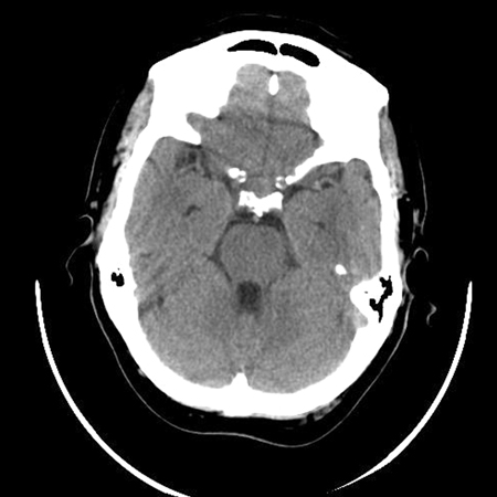
Plain computed tomography scan showing a small pituitary mass encroaching on the right cavernous sinus
BMJ Case Reports 2009; doi:10.1136/bcr.08.2009.2193
See this image in context in the following section/s:
Assessment of pituitary mass
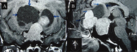
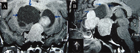
Coronal (A) and sagittal (B) post-contrast magnetic resonance image T1 weighted sections showing a large intrasellar tumour with suprasellar component. Suprasellar solid component shows a lobulated margin with peripheral non-enhancing cystic areas (margins shown by arrows), which are mildly hyperintense compared with cerebrospinal fluid in basal cisterns
BMJ Case Reports 2009; doi:10.1136/bcr.01.2009.1483
See this image in context in the following section/s:
Assessment of pituitary mass
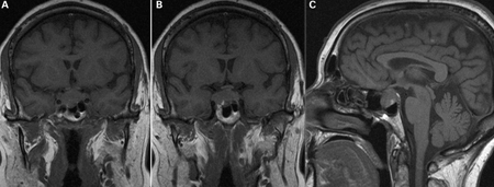
A) Coronal T1 weighted magnetic resonance imaging (MRI) scan showing a pituitary mass with expansion of the pituitary fossa. (B) Coronal T1 weighted MRI scan showing a pituitary mass extending into the cavernous sinus, particularly on the right. (C) Sagittal T1 weighted MRI scan of the pituitary tumour
BMJ Case Reports 2009; doi:10.1136/bcr.08.2009.2193
See this image in context in the following section/s:
Use of this content is subject to our disclaimer