Urgent considerations
See Differentials for more details
Oral cancer
Oral cancer ranks as the sixth most common malignancy worldwide and the third most common cancer in developing countries.[80] Prompt referral for assessment, biopsy, and treatment is mandatory if there is any suspicion of an oral malignancy or for any lesion that does not respond as anticipated within 2 weeks. This is critical, as diagnostic delay increases the risk of the patient ultimately presenting with advanced-staged disease.[85]
Squamous cell carcinoma is the most common form of oral cancer. Recognised contributing factors include tobacco and alcohol use, human papillomavirus infection, immunosuppression, genetic mutations, and diets low in fruit and vegetables.[81][82] Men are affected more frequently than women and the average age of occurrence is >40 years. Oral carcinoma is typically identified in its later stages, and overall 5-year survival rates do not exceed 60%.[81] Early features are non-specific white, red, or red and white mucosal changes with or without ulceration. Symptoms of more advanced disease include bleeding, loosening of the teeth, difficulty wearing dentures, dysphagia, dysarthria, odynophagia, and development of a neck mass.[83] While any site in the mouth may be affected, the most commonly affected areas include the ventrolateral border of the tongue, the floor of the mouth, and the soft palate complex.[83][84][Figure caption and citation for the preceding image starts]: Oral squamous cell carcinomaFrom the collection of Dr Huber [Citation ends].
Malignant salivary gland tumours (mucoepidermoid carcinoma and adenoid cystic carcinoma) are rare and characterised by rapid growth or a sudden growth spurt. They are firm, nodular lesions and can be fixed to adjacent tissue, often with a poorly defined periphery. Pain and neural involvement are common. Eventually, the overlying skin or mucosa may become ulcerated and the adjacent tissues may be invaded. The most common salivary gland tumours that can present as oral ulceration are mucoepidermoid carcinoma and adenoid cystic carcinoma.[2] Surgery, followed by radiotherapy, is the preferred treatment for resectable disease. There is no effective chemotherapy for salivary gland cancer.
Other oral malignancies include oral melanoma and Kaposi's sarcoma, which may manifest as simple ulcerations.[2] Non-Hodgkin's lymphoma may also manifest with oral ulcerations. Oral melanoma is extremely rare, accounting for <1% of primary melanomas. It is characterised by pigmented or amelanotic (white, red, or mucosa-coloured) macular lesions of varying size (1 mm to ≥1 cm), predominantly affecting the palate and maxillary gingiva. It is asymptomatic in the early stages of the disease but may lead to loosening of the teeth, bleeding, ulceration, and pain in advanced stages. Oral lesions in Kaposi's sarcoma may be the initial site of disease in about 15% of patients with AIDS. Kaposi's sarcoma affects the hard palate, gingiva, and dorsum of the tongue and presents as macules, papules, nodules, and exophytic masses of varying size and colour. Advanced lesions may become ulcerated from masticatory trauma and secondary infection.
Stevens-Johnson syndrome and toxic epidermal necrolysis
Erythema multiforme (EM) is a group of diseases caused by an allergic reaction to medications, infections, or illness. They are characterised by mucosal erythema and ulcerations with varying degrees of cutaneous involvement.[37][38][Figure caption and citation for the preceding image starts]: Erythema multiforme in a 53-year-old manFrom the collection of Dr Huber [Citation ends].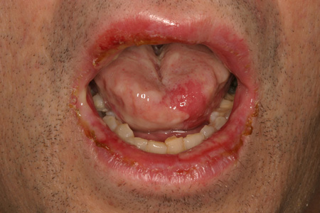 The group includes EM minor, EM major, Stevens-Johnson syndrome (SJS), and toxic epidermal necrolysis (TEN).[39]
SJS and TEN are associated with significant morbidity and potential death.[37][40] Prompt recognition, referral, and management is therefore mandatory. The incidence of TEN is estimated to be 1 to 2 cases per million population per year. The incidence of SJS is estimated to be 1 to 6 cases per million population per year.[37] SJS is a severe form of EM major but is less severe than TEN.
Diagnosis of these conditions is based on the characteristic clinical presentation and skin biopsy results. There is a typical abrupt onset of mouth and lip ulcers with or without cutaneous, ocular, or genital lesions. These lesions may be recurrent and there may be a history of antecedent exposure to drugs, toxins, infection, or immunisation. Constitutional signs and symptoms of pallor, fatigue, malaise, shortness of breath, tachycardia, headache, and irritability may also be present.
SJS is characterised by atypical flat target lesions and multiple involved mucosal sites. TEN presents with extensive cutaneous involvement (10% to ≥30% body surface affected), poorly defined lesions with extensive epidermal detachment, and mucosal lesions similar to those of SJS.
Fungal infection
Mycoses (e.g., zygomycosis, aspergillosis, histoplasmosis, blastomycosis, and paracoccidioidomycosis) associated with the formation of oral ulcers are rare but may be rapidly progressive and potentially fatal.[54][56] Prompt recognition, referral, and treatment is therefore mandatory.
In healthy people, ulcerative oral fungal infections are uncommon, but they may occur in people with uncontrolled diabetes mellitus, haematological malignancy, or immunosuppression (e.g., HIV).[1][54][55][56] The ulcers often are either an extension of a primary lesion in the paranasal sinus or a manifestation of a systemic mycosis. They typically present as deep-seated ulcers, most commonly affecting the tongue, palate, and maxillary alveolar process.[1][54][55] Gingival involvement is uncommon.
In zygomycosis, oral ulcerations, sinusitis, or facial cellulitis may be present. In aspergillosis, yellow or black lesions with a necrotic ulcerated base may be present and are typically located on the palate or posterior tongue. In histoplasmosis, chronic nodular, indurated, or granular masses and ulceration with tissue destruction and bone erosion may be observed. Up to 40% to 50% of patients with systemic histoplasmosis manifest oral lesions, and the major oral sites affected include the mucosa, tongue, palate, and gingiva. In blastomycosis, single or multiple mucosal ulcerations, sessile projections, and granulomatous or verrucous lesions may be present. In paracoccidioidomycosis, oral lesions are common and typically manifest as oral ulcerative granulomas affecting any part of the oral cavity.
Pemphigus
This is a group of autoimmune blistering diseases. Pemphigus vulgaris (PV) and paraneoplastic pemphigus can involve the skin and mucosal surfaces of the mouth, eyes, nasopharynx, and oesophagus. [Figure caption and citation for the preceding image starts]: Pemphigus vulgaris in a 65-year-old womanFrom the collection of Dr Huber [Citation ends].
PV is a rare, potentially life-threatening, intraepithelial splitting disorder with an annual incidence of 1 to 5 cases per million population.[22] It has no sex predilection and the typical age of onset is 40 to 60 years. Ashkenazi Jews and people of Mediterranean origin have a distinct genetic predisposition for PV.[23] About 90% of patients manifest with chronic oral ulcerations, with areas often subjected to trauma (e.g., buccal mucosa, tongue, palate) being the most commonly affected. Oral lesions are the first manifestation of the disease in >50% of people.[24][25] The typical oral lesion appears as a painful erosion/ulcer with an irregular border of necrotic epithelium and is partially covered by a fragile membrane.[2] The Nikolsky sign tends to be positive (i.e., slight rubbing of the skin exfoliates the outermost layer). Concurrent skin or eye lesions, as well as lesions in the nasopharynx and oesophagus, may be present. Two autoantibodies are associated with PV. Anti-desmoglein 1 is associated with cutaneous lesions, and anti-desmoglein 3 is associated with mucosal (oral) lesions.[23]
Paraneoplastic pemphigus is the least common, but most serious, form of pemphigus.[23] Most cases occur in patients who have already developed cancer. Clinical features include acute or chronic mouth sores (flaccid bullae, irregular erosions, and ulcerations) commonly located on the gingiva, buccal mucosa, tongue, and palate; ocular symptoms (conjunctivitis, symblepharon); and concurrent skin lesions.
Use of this content is subject to our disclaimer