Approach
In many patients, oral ulcerative conditions are diagnosed after a careful correlation of the history with clinical findings, and laboratory testing is not required. However, as many oral ulcerative conditions mimic each other, follow-up for the anticipated outcome is essential.
Clinical history
Obtaining a thorough history is essential in assessing a patient presenting with oral ulceration.[86][87] To avoid differential bias, a full history should be obtained before the clinical examination. If an oral ulceration is encountered as part of a physical examination, it is good practice to step back and refocus on the history. Specific areas to address are as follows.
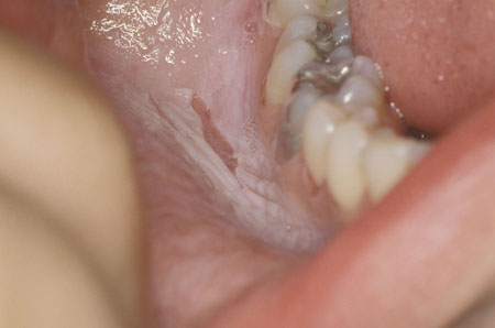
Cause and effect: does the patient suspect or acknowledge a temporally related event or exposure they believe is related to the ulceration? Such findings may be obvious for situations such as a simple cheek bite or pizza burn on the palate (inadvertent trauma) but only suggest suspicion of sensitivity to medications, hygiene products, or foods (e.g., contact stomatitis, erythema multiforme [EM], Stevens-Johnson syndrome [SJS], toxic epidermal necrolysis [TEN], lichenoid reaction). In self-inflicted trauma, patient awareness varies from none, as may occur with encephalitis, to compulsive, as may occur with mental illness.[Figure caption and citation for the preceding image starts]: Burn from topical aspirinFrom the collection of Dr Huber [Citation ends].
 [Figure caption and citation for the preceding image starts]: Oral lichen planusFrom the collection of Dr Huber [Citation ends].
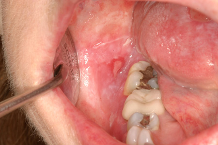
Onset of the complaint: acute onset is most characteristic of ulcers associated with trauma (e.g., iatrogenic trauma from medical or dental procedure), EM, minor recurrent aphthous stomatitis (RAS), and infection. In graft-versus-host disease there may be a history of haematopoietic stem cell transplantation. In ulcers associated with sinus tract, there may be history of dental trauma, infection, or untreated dental disease.
Duration of the complaint: most oral ulcerations resolve within 2 weeks. Any ulceration that does not improve within 2 weeks should be considered for biopsy. Conditions associated with oral ulceration chronicity include malignancy, nutritional deficiencies (e.g., iron, folate, vitamin B12, or vitamin C deficiencies), and several dermatological, immunological, and infectious conditions (e.g., RAS, lichen planus, pemphigus, mucous membrane pemphigoid [MMP], and fungal infections). Oral cancer ranks as the sixth most common malignancy worldwide and the third most common cancer in developing countries.[80] Prompt referral for assessment, biopsy, and treatment is mandatory if there is any suspicion of an oral malignancy or for any lesion that does not respond as anticipated within 2 weeks. This is critical, as diagnostic delay increases the risk of the patient ultimately presenting with advanced-staged disease.[85][Figure caption and citation for the preceding image starts]: Pemphigus vulgaris in a 65-year-old womanFrom the collection of Dr Huber [Citation ends].
 [Figure caption and citation for the preceding image starts]: Mucous membrane pemphigoid in a 53-year-old womanFrom the collection of Dr Huber [Citation ends].
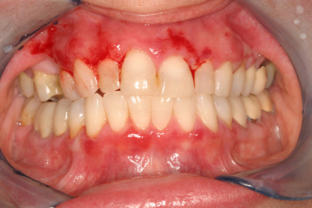
Recurrence: a history of prior occurrence is noteworthy. Notable recurrent conditions include RAS and recurrent herpes. Continued exposure to a known or unknown allergen or trigger may result in recurrence of EM, contact stomatitis, and lichenoid reactions.[Figure caption and citation for the preceding image starts]: Erythema multiforme in a 53-year-old manFrom the collection of Dr Huber [Citation ends].
Prodrome: some patients relate an awareness (e.g., altered sensation, swelling) of impending RAS or recurrent herpes ulceration.
Behavioural habits and practices: high-risk behaviours such as drug and alcohol abuse, tobacco use, and unprotected sex are associated with increased risk of STDs (syphilis, gonorrhoea) and oral cancer (squamous cell carcinoma). For ulcers associated with tuberculosis there may be a history of homelessness or institutionalisation.
Extra-oral involvement: oral ulcerations often are one component of a more global condition (e.g., EM, lichen planus, chronic ulcerative stomatitis, pemphigus, MMP, linear IgA bullous dermatosis, epidermolysis bullosa acquisita, periodic fever syndromes, reactive arthritis, lupus erythematosus, giant cell arteritis, granulomatosis with polyangiitis (formerly Wegener's granulomatosis), Behcet's disease, nutritional deficiency, infections). Of note is the fact that the oral ulceration may be the first manifestation (herald lesion) of the underlying disease. For ulcers associated with nutritional deficiencies, there may be history of anaemia, dieting, alcoholism, absorptive disorders (e.g., Crohn's disease, coeliac disease, ulcerative colitis). In addition, the patient may have constitutional signs and symptoms of pallor, fatigue, and malaise.
Comprehensive systems review: numerous systemic conditions (e.g., diabetes mellitus, gastrointestinal diseases, liver diseases, malnutrition, malignancy, or alcoholism) may underlie an oral ulceration. As a rule, conditions of impaired immunocompetence must be carefully considered. For immunocompromised patients who present with an oral ulceration, the potential aetiologies expand dramatically, and the clinical presentation may be more severe and widespread. These patients require a more aggressive and comprehensive assessment. Several fungal infections (zygomycosis, aspergillosis, histoplasmosis, blastomycosis) that cause ulcers are associated with immunosuppressive states.
Pain and impact on function: while most oral ulcerative conditions manifest some degree of discomfort, it may be useful to have patients score their pain and describe any impact on function (e.g., eating, drinking, swallowing, speaking). As an example, an inability to maintain hydration and nutrition while having primary herpetic gingivostomatitis increases the patient's risk for dehydration and secondary infection. Acute or chronic mouth pain (particularly on eating and drinking) is often associated with lichen planus and chronic ulcerative stomatitis. Swallowing impairment or altered sensation associated with a long-standing ulcerative nodule on the posterior lateral border of the tongue is characteristic of locally advanced carcinoma.
Physical examination
Examination of the patient should be thorough and disciplined.[87] A complete visual and tactile assessment of the soft tissues of the head and neck should be accomplished before focusing on the chief complaint.[88] Factors to consider include the following.
Fever: this suggests an infectious or possible immunological aetiology (e.g., periodic fever syndromes).
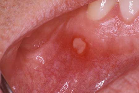
Location of ulcerative lesions: lesions restricted or prone to certain locations tend to be associated with specific conditions. For example, in RAS, the keratinised surface is spared; in herpangina, ulcers are localised to the soft palate or tonsil pillars; in recurrent herpes, ulcers are restricted to the keratinised surfaces; and in MMP, the gingiva may be involved. In nutritional deficiencies, ulcers may affect the tongue (glossitis and angular cheilitis). In granulomatosis with polyangiitis (formerly Wegener's granulomatosis) there may be hyperplastic petechiae-laden (strawberry) gingivitis. In necrotising ulcerative gingivitis, the bacterial infection typically affects the interdental and marginal gingival tissues.[Figure caption and citation for the preceding image starts]: Aphthous ulcerFrom the collection of Dr Huber [Citation ends].
 [Figure caption and citation for the preceding image starts]: Mucous membrane pemphigoid in a 53-year-old womanFrom the collection of Dr Huber [Citation ends].[Figure caption and citation for the preceding image starts]: Necrotising ulcerative gingivitisFrom the collection of Dr Huber [Citation ends].
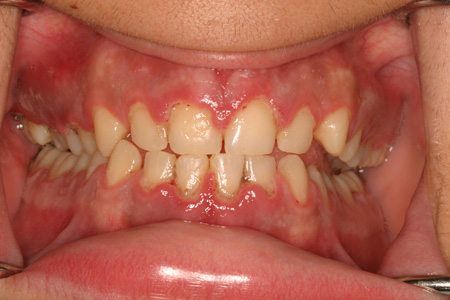 [Figure caption and citation for the preceding image starts]: Primary herpetic gingivostomatitis in a 15-year-old girlFrom the collection of Dr Huber [Citation ends].
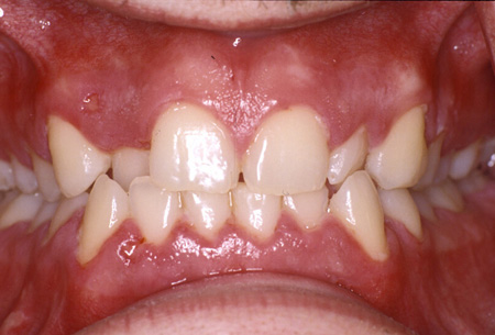
Size of ulcerative lesions: lesions of viral origin tend to initially be small (1-2 mm in diameter).
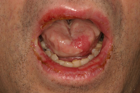
Shape of ulcerative lesions: oval or round lesions are characteristic of RAS, Behcet's disease, and initial viral eruptions. Irregularly shaped lesions suggest lichen planus, pemphigus, MMP, major RAS, or carcinoma. As viral lesions (e.g., herpetic ulcers) develop, individual lesions can coalesce to form an irregularly shaped ulcer. Long-standing oral ulcers often have a raised keratotic border. In lichen planus and oral lichenoid reaction, lacey white striations (Wickham's striae) are usually present. In lupus erythematosus, there may be fine stippling of white dots on the ulcers.[Figure caption and citation for the preceding image starts]: Oral squamous cell carcinomaFrom the collection of Dr Huber [Citation ends].
 [Figure caption and citation for the preceding image starts]: Aphthous ulcerFrom the collection of Dr Huber [Citation ends].[Figure caption and citation for the preceding image starts]: Oral lichen planusFrom the collection of Dr Huber [Citation ends].[Figure caption and citation for the preceding image starts]: Pemphigus vulgaris in a 65-year-old womanFrom the collection of Dr Huber [Citation ends].[Figure caption and citation for the preceding image starts]: Mouth ulcer in Behcet's syndromeWith kind permission of Dr Y. Yazici [Citation ends].
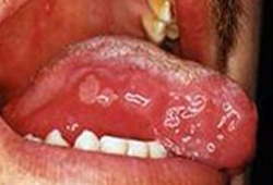
Induration of ulcerative lesion: this suggests infection (e.g., fungal, syphilitic chancre) or carcinoma. Extensive deep ulcers with indurated borders located in hard or soft palate are characteristic of necrotising sialometaplasia.
Lymphadenopathy: tender, smooth, freely moveable nodes suggest acute inflammatory disease. Matted, non-tender, firm, or rubbery nodes suggest malignancy.
Extra-oral involvement: extra-oral manifestations may be present, such as target lesions of EM, cutaneous blisters associated with pemphigus, a cutaneous eruption on hands and feet in hand-foot-and-mouth disease, and conjunctivitis in Behcet's disease, MMP, EM, linear IgA bullous dermatosis, and epidermolysis bullosa acquisita. The classic cutaneous lesion of lichen planus presents as a purple, polygonal papule.[20] Nail manifestations are uncommon and consist of thinning and ridging of the nail plate with splitting of the distal free edge.[20]
The Nikolsky sign tends to be positive (i.e., slight rubbing of the skin exfoliates the outermost layer) in several lesions including pemphigus, MMP, paraneoplastic pemphigus, oral lichen planus, epidermolysis bullosa, linear IgA disease, EM, and graft-versus-host disease.[23]
Laboratory tests
Correlating the history with findings from the physical examination is often all that is necessary to determine the correct diagnosis and initiate appropriate management. However, when the oral ulceration does not respond as anticipated to treatment or the initial presentation is equivocal, further laboratory testing or referral is required. This may include the following.
Biopsy for histopathology: this is the cornerstone of diagnosis for any chronic oral ulceration.[81] Direct immunofluorescence is often used to assess lesions where the diagnosis is not entirely clear after clinical examination and standard histopathology. If the primary clinician is not comfortable performing the biopsy, a referral is warranted. In general, the biopsy specimen should include a portion of clinically normal tissue. This is especially important if immunofluorescence testing is indicated, because the specimen undergoing testing must be intact. While a specimen obtained from the centre of an ulcer may prove sufficient to diagnose some conditions (e.g., deep fungal infection, oral carcinoma), there is a concern that such a specimen may harvest only necrotic non-diagnostic tissue.
Blood and/or chemistry testing: this is indicated when an underlying systemic condition such as malnourishment or infection is suspected. It generally consists of an FBC and peripheral blood smear, haematinic screen (serum iron, ferritin, folate, and vitamin B12), liver function tests, and erythrocyte sedimentation rate. Blood tests may be useful for excluding possible haematological disorders, HIV infection, or diabetes mellitus. More targeted testing is dictated by the clinical impression.[62]
Microbiological and serological tests: these are undertaken when an infectious agent is suspected - for example, to determine a fungal infection (zygomycosis, aspergillosis, histoplasmosis, blastomycosis, paracoccidioidomycosis). They may also be used for Sm antigen in lupus erythematosus, HLA-B27 in reactive arthritis (Reiter's syndrome), antineutrophil cytoplasmic antibody (ANCA) in granulomatosis with polyangiitis (formerly Wegener's granulomatosis), and serum treponemal enzyme immunoassay in syphilis. It should be noted that public health protocols recommend universal screening of all pregnant women for syphilis.[89][90]
Imaging: this may be used to ascertain the involvement of adjacent structures. For example, a sinus x-ray and intra-oral periapical x-ray may be used when the suspected cause of ulceration is sinus tract disease.
Referral to specialist
Some patients may require referral to an oral medicine specialist or an oral and maxillofacial surgeon. Referral may be indicated for a variety of reasons:[87]
The diagnosis is uncertain
The lesion is not responding to treatment
The management of the lesion is not within the scope of the primary clinician
The patient requests a second opinion
The ability of the patient to tolerate or undergo the indicated therapy is in doubt
The primary clinician believes a specialist is better equipped or prepared to manage the lesion.
Use of this content is subject to our disclaimer