Images and videos
Images


Assessment of seronegative arthritis
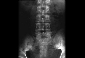
Plain radiograph showing bilateral sacroiliitis in a patient with ankylosing spondylitis
BMJ 2006;333:581-585. Copyright@2009 by the BMJ Publishing Group
See this image in context in the following section/s:
Assessment of seronegative arthritis
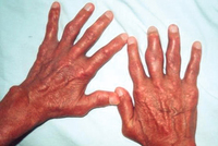
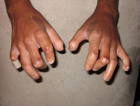
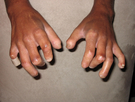
Late-stage sclerodactyly with contractures due to severe skin tightening
From the collection of Maureen D. Mayes; used with permission
See this image in context in the following section/s:
Assessment of seronegative arthritis
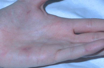
Janeway lesions
From the collection of Sanjay Sharma; used with permission
See this image in context in the following section/s:
Assessment of seronegative arthritis
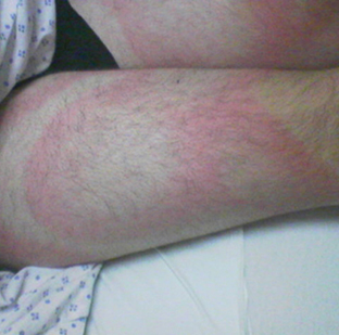
Erythema migrans
From the collection of Dr Cristian Speil; used with permission
See this image in context in the following section/s:
Assessment of seronegative arthritis
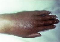
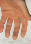
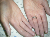
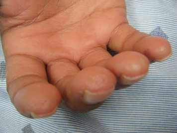
Hand demonstrating sclerodactyly with finger curling, shiny skin at the fingers, and telangiectasias
From the collection of Maureen D. Mayes; used with permission
See this image in context in the following section/s:

Assessment of seronegative arthritis
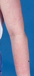
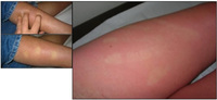
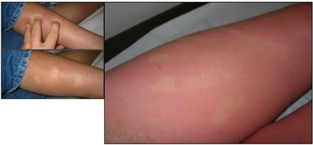
Lacy, reticular, erythematous eruption of erythema infectiosum on an upper extremity
From the collection of Gary A. Dyer, MD; used with permission
See this image in context in the following section/s:

Assessment of seronegative arthritis
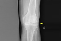
Knee radiograph with linear calcific deposits of cartilage calcification
From the collection of Ann K. Rosenthal, MD; used with permission
See this image in context in the following section/s:

Assessment of seronegative arthritis
Erythema nodosum on the shin of a patient
From the collection of Dr Om P. Sharma; used with permission
See this image in context in the following section/s:

Assessment of seronegative arthritis
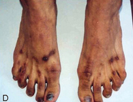
Macular erythematous patches (Gottron's papules) over the toes with dystrophic cuticles
Adapted from BMJ Case Reports 2009 [doi:10.1136/bcr.06.2009.2027] Copyright © 2009 by the BMJ Publishing Group Ltd
See this image in context in the following section/s:
Assessment of seronegative arthritis
Psoriatic arthritis
From the collection of Dr Tsu-Yi Chuang; used with permission
See this image in context in the following section/s:

Assessment of seronegative arthritis
Chronic tophaceous gout showing nodules in periarticular structures and arthritis
Adapted from BMJ Case Reports 2009 [doi:10.1136/bcr.03.2009.1668] Copyright © 2009 by the BMJ Publishing Group Ltd
See this image in context in the following section/s:

Assessment of seronegative arthritis
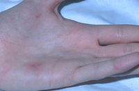
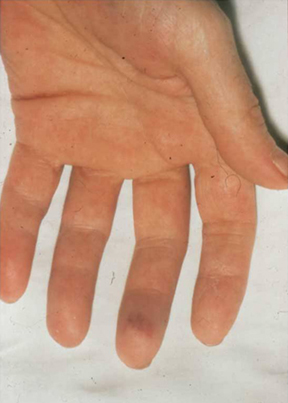
Hands demonstrating Raynaud's phenomenon
From the collection of Maureen D. Mayes; used with permission
See this image in context in the following section/s:

Assessment of seronegative arthritis
Cutaneous infarcts
From the collection of Sanjay Sharma; used with permission
See this image in context in the following section/s:
Assessment of seronegative arthritis
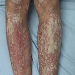
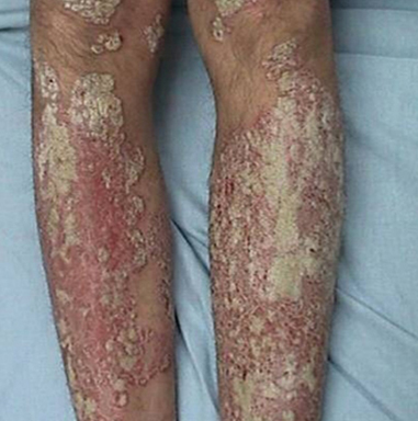
Psoriasis plaque: legs
From the collection of Dr Tsu-Yi Chuang; used with permission
See this image in context in the following section/s:

Assessment of seronegative arthritis
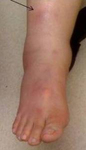
Gonococcal arthritis of the hand, which caused the hand and wrist to swell
CDC/ Susan Lindsley, VD; used with permission
See this image in context in the following section/s:
Assessment of seronegative arthritis
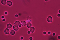
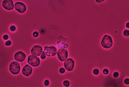
Image of intracellular calcium pyrophosphate crystals under compensated polarising light microscopy
From the collection of Ann K. Rosenthal, MD; used with permission
See this image in context in the following section/s:
Assessment of seronegative arthritis
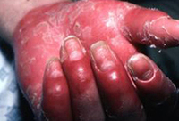
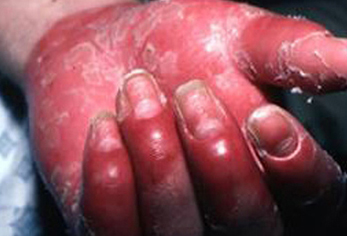
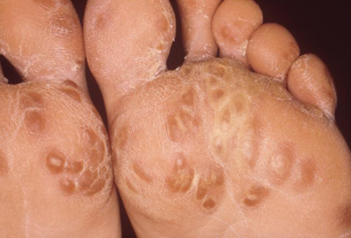
Keratoderma blennorrhagica in a patient with reactive arthritis
Image provided by the CDC Public Health Image Library
See this image in context in the following section/s:
Assessment of seronegative arthritis
Osler's node
From the collection of Sanjay Sharma; used with permission
See this image in context in the following section/s:

Assessment of seronegative arthritis
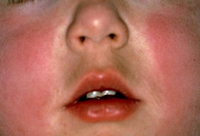
Typical erythematous 'slapped cheeks' of erythema infectiosum
From the collection of Gary A. Dyer, MD; used with permission
See this image in context in the following section/s:
Assessment of seronegative arthritis
Features of spondyloarthropathies
Adapted from Kataria RK, Brent LH. Am Fam Physician. 2004;69:2853-2260 and Gladman DD. Am J Med Sci.1998;316:234-238
See this image in context in the following section/s:
Assessment of seronegative arthritis
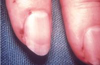
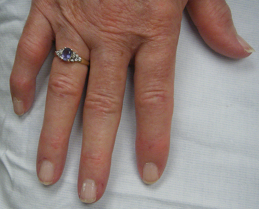
Digital pits without ulcers
From the collection of Maureen D. Mayes; used with permission
See this image in context in the following section/s:

Assessment of seronegative arthritis
Discriminating features in polyarthritis
Adapted from Pinals R. N Engl J Med. 1994;330:769-774
See this image in context in the following section/s:
Assessment of seronegative arthritis
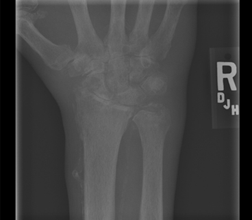
Wrist radiograph from a patient with chronic calcium pyrophosphate arthritis showing severe degenerative changes
From the collection of Ann K. Rosenthal, MD; used with permission
See this image in context in the following section/s:
Assessment of seronegative arthritis
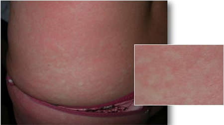
Diffuse hyperaemia or erythroderma during the acute phase of chikungunya virus infection
From the collection of Dr Fabrice Simon; used with permission
See this image in context in the following section/s:

Assessment of seronegative arthritis
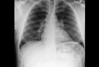
Bilateral hilar adenopathy
From the collection of Muthiah P. Muthiah, MD, FCCP; used with permission
See this image in context in the following section/s:
Assessment of seronegative arthritis
Diffuse morbilliform rash during the acute phase of chikungunya virus infection
From the collection of Dr Fabrice Simon; used with permission
See this image in context in the following section/s:
Use of this content is subject to our disclaimer