Approach
Clinical assessment helps determine the impact of the patient's snoring, helps decide whether the patient needs a sleep study to rule out obstructive sleep apnoea (OSA), and helps identify whereabouts in the upper airway the snoring is originating from (which helps target treatment).
Unfortunately, history and clinical findings have been shown to lack sensitivity and specificity for diagnosing OSA. It is estimated that history and examination can only predict OSA in 50% of patients, although male sex, snoring, and increased BMI have been found to be useful predictors of OSA.[24][25]
For more information on obstructive sleep apnoea, see the BMJ Best Practice topics Obstructive sleep apnoea in adults and Dyssomnias in children.
History
It is sometimes helpful to have the patient's partner present during some or all of the consultation to provide information on some features of the snoring episodes. It is important to ascertain how long the patient has been snoring for and if the snoring is disturbing the patient, the partner, or both. Severity of snoring can be assessed by asking if the snoring can be heard by the next-door neighbour, anywhere in the house, in an adjacent room, or just in the same room. The level of disturbance caused by snoring can be indicated by whether the partner sleeps separately and, if so, how often. It is also important to determine if the snoring is worse, or only occurs, when the patient is lying on their back.
Features that may suggest OSA should be sought:[1]
Witnessed apnoeas (i.e., the patient stops breathing for 10 seconds or more)
Choking or gasping episodes
Waking up tired, with a headache
Daytime somnolence
Past medical history of diabetes, hypertension, or ischaemic heart disease, particularly in relatively young patients.
Patients should be asked whether they have a history of blocked nose and whether it affects the left side, right side, or both sides. Nasal allergies such as hay fever can contribute to snoring.[1] The patient should be asked about any pets, as patients may be allergic to them.
Smoking habit and alcohol intake should also be noted. Smoking causes inflammation and congestion of the upper airway.[5][20][21] Active and passive smoking are risk factors for snoring.[1] The frequency of habitual snoring increases with the amount of tobacco smoked.[20] Alcohol causes a deeper sleep and lowering of upper airway muscle tone.[1][15][16][17] Alcohol dependence has been associated with self-reported snoring in lean women.[18]
Medication history may identify drugs that may contribute to snoring. Sedating medications such as sleeping tablets, tranquillisers, and antihistamines lower upper airway muscle tone and predispose to snoring.[1][11]
The patient should be asked what treatments or techniques they have already tried and how successful they have been, in order to avoid repetition.
Enquiry about any changes in the patient's weight should be made. The amount of weight gained and the timescale should be quantified. If the patient did not snore before gaining weight, then weight loss may be curative.
In children, heavy snoring with disturbed sleep, respiratory pauses, and snort arousal (briefly wakening after snorting) suggests OSA. Night terrors, enuresis, hyperactivity, and behaviour problems are also associated with OSA.
Patient questionnaires
There are a number of patient questionnaires available that can be useful in evaluating a patient who snores. The snoring scale score is a patient questionnaire that measures the severity of snoring by asking three questions looking at the loudness, frequency, and periodicity of the snoring, with four possible responses (0 to 3), giving a possible maximum score out of 9.[26]
A measurement of excessive daytime somnolence (EDS) may subjectively be made by the Epworth sleepiness score (ESS) patient questionnaire. Epworth Sleepiness Scale Opens in new window Patients are asked to rate on a scale of 0 to 3 the likelihood of falling asleep in 8 specific situations. A score above 10 (out of 24) indicates EDS. However, the ESS lacks sensitivity and specificity as a screening tool for OSA, so in an individual patient the ESS may not on its own be very useful and only acts as a guide.[12]
Examination
The following features, in particular, should be assessed during physical examination of a patient who snores.
General examination
The clinical appearance may suggest that a patient has hypothyroidism or acromegaly.
BMI
BMI is a measurement that helps define the degree of obesity. It is calculated by dividing the weight in kilograms by the height in metres squared (kg/m²). Normal BMI = 19 to 25, overweight = 26 to 30, Class I obesity = 30 to 34.9, Class II obesity = 35 to 39.9, Class III obesity = >40.
Cranio-facial abnormalities
Underlying skeletal anatomy may result in a narrowed airway predisposing to snoring. Retrognathia or micrognathia prevents the tongue from being positioned sufficiently forwards during sleep, thereby also predisposing to snoring.
Nose
External nasal deformities may contribute to restricted nasal airflow. For example, deviated nasal bones often indicate a deviated nasal septum with reduced nasal airflow.
The patient should be asked to inhale through the nose and see if there is any alar collapse (i.e., inwards movement of the outside of the nostrils).
The inside of the nose should be examined with an otoscope, a headlight, or, ideally, a nasendoscope.
 How to examine the nasal cavity
Video outlining how to perform an examination of the nose and nasal cavity
Large, pale inferior turbinates are associated with allergic rhinitis.
There may be turbinate hypertrophy, a deviated nasal septum, nasal polyps, or, rarely, a tumour causing nasal obstruction.
Tongue size
Looking into the mouth with the tongue at rest, the tongue size can be graded 1 to 4 according to the modified Mallampatti score, where 1 = can visualise the tonsils, 2 = can visualise the uvula, 3 = can visualise the soft palate, 4 = can visualise only the hard palate.
Tonsil size
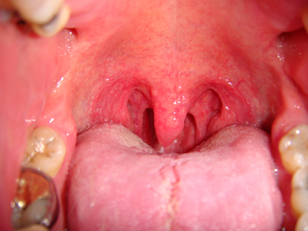
Looking into the mouth, the tonsil size can be graded 0 to 4, where 0 = no tonsils present, 1 = tonsils within the pillars, 2 = tonsils up to the edge of the pillars, 3 = tonsils beyond the pillars, 4 = tonsils extend to the midline.[Figure caption and citation for the preceding image starts]: Crowded oropharynx due to tonsils, prominent posterior pillars, and large uvulaFrom the collection of Dr Showkat Mirza; used with permission [Citation ends].
Soft palate
Soft palate anatomy is highly variable and some features may contribute to snoring, such as the presence of posterior pillar and uvular webbing, and an excessively long soft palate and uvula.
Upper airway
Flexible endoscopy allows a complete assessment of the upper airway, from the nasal cavity to the larynx, looking for predisposing factors for snoring such as adenoidal tissue, lingual tonsils, abnormalities of the epiglottis, and, occasionally, hypo-pharyngeal cysts or tumours. Adenoidal tissue is a common cause of snoring in children with or without tonsillar hypertrophy. Adenoid hypertrophy is rare in adults, but when it does occur it is usually in young adults.
Müller manoeuvre
This technique may differentiate between palatal and tongue base or multi-segmental obstruction. Flexible nasendoscopy is performed with the patient in the sitting position and with the mouth closed. The fibre-optic endoscope is positioned at the level of the tongue base. The patient inhales vigorously while the nares and mouth are occluded and the degree of hypo-pharyngeal collapse noted. This manoeuvre is then repeated with the endoscope positioned just above the soft palate (velopharyngeal level).
This may help assess the suitability of patients for uvulopalatopharyngoplasty (UPPP).[27]
Passive endoscopic hypotonic method
This is another technique that may differentiate between palatal and tongue base or multi-segmental obstruction. Flexible nasendoscopy is performed in the awake patient in the supine position at end expiration (i.e., the patient exhales maximally) when phasic muscle tone is at its lowest, and this may point to where the obstruction is occurring during sleep.[28]
Tests and investigations
Depending on the findings of the history and examination, a number of further tests and investigations may be indicated.
If suspected clinically, TFTs may confirm or exclude hypothyroidism as a contributory cause to snoring. Similarly, if there is a clinical suspicion of acromegaly, measurement of growth hormone levels can provide supporting evidence. If a patient has allergic rhinitis, skin-prick tests and serum allergen-specific IgE tests can identify specific allergens. If nasal obstruction is thought to be a significant contributory cause, a nasal decongestant test can be useful. This involves using a topical nasal decongestant on alternate nights over a 1-week period and comparing the severity of snoring. If the decongestant results in improved symptoms, it may be worth treating nasal abnormalities to improve the snoring.[29]
An overnight sleep study may be used if there is concern over possible OSA. The simplest sleep study is overnight pulse oximetry. This measures oxygen saturation and provides pulse rate data. It assumes that when an individual has an apnoeic or hypopnoeic episode, the oxygen saturation falls. Once the apnoea or hypopnoea is relieved, the oxygen desaturation recovers. The falls and rises are regarded as oxygen dips.[12] Some clinicians use oximetry alone as a screen for OSA. Unfortunately, studies using overnight oximetry as a screening tool for OSA have shown good specificity and positive predictive value, but poor sensitivity and negative predictive value.[30] This means overnight oximetry may miss subjects with OSA who do not desaturate. It may also miss patients with upper airway resistance syndrome (UARS). Full polysomnography remains the definitive method for diagnosing OSA.[12] An apnoea-hypopnoea index (AHI) of over 5 indicates OSA.
Pharyngeal manometry, in which pressure probes are inserted per nasally into the upper airway, with an overnight sleep study, may help identify whether the snoring is occurring at the palate or tongue base level.[31] This is usually only done in patients who have expressed a wish to be considered for palate surgery and are a suitable candidate.
Sleep nasendoscopy may help identify the site of origin of snoring and select patients suitable for palate surgery.[32] It involves sedating the patient with propofol to a level of sleep sufficient to induce snoring. With the patient in the supine position, the operator then examines the upper aerodigestive tract with a flexible nasendoscope to determine the level(s) of obstruction. Again, this is usually only done in patients who have expressed a wish to be considered for palate surgery and are suitable candidates.
Imaging studies may also have a useful role in identifying sites of narrowing and obstruction. Three-dimensional CT of the head and neck may be useful to evaluate upper airway patency in patients with sleep-disordered breathing.[12] Ultrafast MRI can be used in awake and asleep patients to assess the site of upper airway obstruction, but it is not available at all centres, and short periods of sleep in an MRI machine may not be representative of normal sleep patterns.[12]
Acoustic analysis of snoring can be helpful in objectively determining the volume and duration of snoring but is only of limited use in detecting the site of snoring.[33] It may also be a useful research tool to evaluate the impact of various environmental and patient factors on the severity of snoring.[34][35]
Use of this content is subject to our disclaimer