Approach
When gangrene is suspected, the most important decision is to differentiate ischaemic gangrene from infectious gangrene and to recognise deep necrotising infections that require urgent surgical intervention.
This usually requires:
Identification of any risk factors
A careful history and comprehensive physical examination
Appropriate laboratory investigations
Imaging and other studies, in selected cases.
Rapid progression, spread, or clinical deterioration indicates a need for surgical exploration. In ambiguous cases, biopsy or surgical exploration is necessary to determine if the fascia is involved, because observation of the deeper soft tissue is the only definitive method to make the diagnosis.[39]
Identification of risk factors
Risk factors for ischaemic gangrene include diabetes mellitus, atherosclerosis, smoking, prolonged application of tourniquets, hypercoagulable states, drug abuse, malignancy, and renal disease.
Risk factors for infectious gangrene include recent trauma or abdominal surgery, contaminated wounds, diabetes mellitus, alcoholism, renal disease, drug abuse, varicella lesion, skin/insect bite, malignancy, immunosuppression, and malnutrition.
Clinical evaluation
Findings suggestive of arterial ischaemic gangrene include: cold extremity, history of previous thrombosis, chronic course, trophic skin changes, pain (classically described as a burning pain in the ball of the foot and toes that is worse at night), purpura, oedema, ulcers, and livedo reticularis.[20]
Blood pressure evaluation is useful in evaluating ischaemic gangrene. An ankle systolic pressure of 50 mmHg or less, or a toe systolic pressure of 30 mmHg or less, supports diagnosis of ischaemic gangrene; however, these parameters may be less reliable in patients with diabetes because arterial wall calcification can impair compression of vessels by a blood pressure cuff and produce systolic pressure measurements that are greater than true levels.[9] Another useful measure is the ankle-brachial index (ABI), which is the ratio of the systolic pressure at the dorsalis pedis or posterior tibial artery divided by the systolic pressure at the brachial artery. Patients with critical limb ischaemia usually have an ABI of 0.4 or less.[9]
Clinically, features that may help categorise the viability of an acutely ischaemic limb include:
Viable: not immediately threatened; no sensory loss or muscle weakness; audible arterial and venous Doppler signals
Threatened marginally: salvageable if promptly treated; no or minimal (toes) sensory loss; no muscle weakness; often inaudible arterial Doppler signals; audible venous Doppler signals.
Threatened immediately: salvageable with immediate revascularisation; sensory loss of more than toes accompanied by rest pain; mild to moderate muscle weakness; usually inaudible arterial Doppler signals; audible venous Doppler signals.
Irreversible/non-viable: major tissue loss and/or permanent nerve damage; profound anaesthesia; profound paralysis (rigor); inaudible arterial and venous Doppler signals.
Phlegmasia cerulea dolens, usually associated with malignancy, causes total or near-total obstruction of venous drainage from a limb. Pain, marked oedema of the limb, and cyanosis of the skin ensue over a variable time course. In approximately one half of cases, the cyanosis may be preceded by a white appearance of the skin, caused by subcutaneous oedema without venous congestion.
Findings suggestive of infectious gangrene include: low-grade fever and chills, purple or red bullous lesions, severe pain on palpation over contiguous but seemingly unaffected areas, indistinct margins, crepitus, loss of sensation distal to the affected area, and rapid progression of the infection.[6]
Necrotising cellulitis (NC) presents with thin dark wound drainage and gas formation in the skin with sparing of fascia and deep muscles. Onset is gradual with no pain, swelling, and systemic toxicity.[40][41]
Necrotising fasciitis (NF) is a deep infection of the subcutaneous tissue that results in progressive destruction of fascia and fat. It is usually erythematous without sharp margins, swollen, warm, shiny, and exquisitely tender with rapid progression over days with changes in skin colour and bullae formation. Frank cutaneous gangrene can be observed in 3 to 5 days with a prodrome of anaesthesia before the appearance of skin necrosis. This helps to distinguish from NC above.[40][41]
Necrotising myositis (spontaneous gangrenous myositis) is an aggressive infection with fever, exquisite pain, and muscle swelling. Overlying skin can transition to erythema, warmth, petechiae, bullae, and vesicles with progression over several hours to involve contiguous muscle groups and soft tissue. Septic shock in the form of streptococcal toxic shock syndrome can occur. Timeline of progression aids in differentiation from NF.[40][41][Figure caption and citation for the preceding image starts]: Haemorrhagic blister formation secondary to ischaemic gangreneCollection of Jose Contreras-Ruiz (Clinica del Cuidado Integral de Heridas y Estomas) [Citation ends]. [Figure caption and citation for the preceding image starts]: Newborn with purpura fulminans due to Streptococcus B haemolyticusCollection of Jose Contreras-Ruiz (Clinica del Cuidado Integral de Heridas y Estomas) [Citation ends].
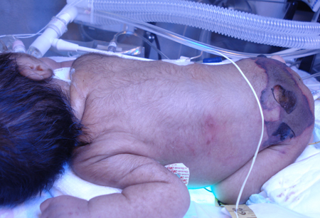
Laboratory testing
For ischaemic and infectious gangrene, initial laboratory assessment includes complete blood count with peripheral blood smear, liver function tests, renal function tests, C-reactive protein (CRP), serum lactate dehydrogenase (LDH), and baseline coagulation studies.
Findings that would be supportive of diagnosis of gangrene and may warrant further investigation include:
CRP >3.26 mmol/L (>150 mg/L)
White blood cell (WBC) count >15 x 10⁹/L (>15,000 cells/microlitre); note that despite serious infection, WBC may not be raised
Haemoglobin <135g/L (<13.5 g/dL); haemolytic anaemia is a common finding in gas gangrene
Sodium <135 mmol/L (<135 mEq/L)
Elevated LDH levels
Lab results suggesting acute renal failure, hepatic failure, or metabolic acidosis.
Blood cultures are recommended if infectious gangrene is suspected.
The LRINEC scoring system is a useful tool to utilise the initial laboratory test to predict the risk of necrotising fasciitis (NF).[3][42] It has a 92% positive predictive value and 96% negative predictive value. Scores are given for levels of CRP, WBC count, haemoglobin, sodium, creatinine, and glucose. Any score of 6 or greater is suspicious for NF, and a score of 8 or greater is highly predictive of NF.
If routine tests are unrevealing, further tests may help diagnose conditions that may contribute to produce ischaemia and gangrene: antinuclear antibodies (Sjogren syndrome, systemic lupus erythematosus, discoid lupus), antiphospholipid antibodies (antiphospholipid syndrome), cold agglutinins (primary or secondary cold agglutinin disease), cryofibrinogens (essential cryofibrinogenaemia or in association with infections, rheumatological disorders, or cancer), and cryoglobulin (associated with lymphoproliferative, inflammatory or infective disease) may all be indicated in patients with suspected ischaemic gangrene causing blue or purple toes.[20]
Gram stain of infected tissue may demonstrate gram-positive bacilli. Small chains of gram-positive cocci suggest a streptococcal infection; clumps of large cocci suggest Staphylococcus aureus.[1] Blood and tissue cultures can provide definitive bacteriological diagnosis and indicate whether infection is polymicrobial or monomicrobial.
Imaging
If a patient has suspected gas gangrene, plain x-rays may demonstrate gas in the soft tissues and/or indicate underlying osteomyelitis. Magnetic resonance imaging (MRI) or computed tomography (CT) scanning may reveal abscess formation or evidence of enhancement, oedema, or thickening in the fascia. However, a lack of demonstrable gas in the soft tissue does not exclude diagnosis of a necrotising infection. CT can reveal smaller collections of gas in the tissues than plain x-rays and so may be preferable in this regard. CT scans can also more readily demonstrate fluid collections. MRI, while offering greater soft-tissue details than CT, is generally less readily available and can also present logistical problems in patients who are often quite severely ill.
In a patient with suspected ischaemic gangrene a number of imaging techniques may be indicated, depending on the exact clinical situation:
Duplex ultrasound can be useful to demonstrate location and severity of arterial narrowing or obstruction and can also detect venous thromboembolism.[20] It is the most widely used modality to assess location and degree of stenosis in peripheral vascular disease as well as patency of bypass grafts. The sensitivity and specificity of 50% or greater stenosis from the iliac artery to popliteal artery are 90% and 95%, respectively.
Conventional angiography is frequently used to identify the site and extent of arterial or venous obstruction. Accuracy and interpretation is enhanced by using techniques such as digital subtraction techniques, which eliminate bony and dense body tissue artifacts. However, it is an invasive procedure requiring contrast.
If available, CT angiography or magnetic resonance angiography (MRA) may be used to look for the presence of atheroemboli. CT angiography is increasingly used, but it still requires intravenous contrast, although there is less radiation than with traditional angiography. It can also reconstruct the images into 3D images. The new 64-slice CT images can have sensitivity from 89% to 100% and specificity from 92% to 100% for >50% stenosis. However, its spatial resolution is lower than digital subtraction angiography and venous opacification can obscure arterial filling. Less widely available, the sensitivity and specificity of MRA to detect a stenosis greater than 50% can be as high as 90% to 100% with the greatest accuracy when gadolinium is used. However, it does have several limitations. MRA tends to overestimate stenosis and occlusions; metal clips can mimic occlusions thus limiting its use in post-surgical patients. Also, patients with pacemakers, defibrillators, and some cerebral aneurysm clips cannot be scanned safely, and gadolinium has caused nephrogenic systemic fibrosis (NSF) in patients with chronic renal insufficiency.
CT scans of the chest and abdomen are indicated in patients where it is suspected that there may be an occult malignancy causing a paraneoplastic thromboembolism syndrome as a cause of ischaemia.
Echocardiography is indicated in cases where features of the history and clinical examination (e.g., history of valvular disease, presence of cardiac murmurs) suggest possible suspected infective endocarditis, myxoma, and non-bacterial thrombotic endocarditis.
Use of this content is subject to our disclaimer