Clinical presentation
Travel history is essential in determining risk of exposure to infection. Most patients in the US have recently returned from a vacation at a beach destination in the tropics or subtropics, especially the Caribbean, Brazil, Mexico, and Southeast Asia.[4]Davies HD, Sakuls P, Keystone JS. Creeping eruption. A review of clinical presentation and management of 60 cases presenting to a tropical disease unit. Arch Dermatol. 1993 May;129(5):588-91.
http://www.ncbi.nlm.nih.gov/pubmed/8481019?tool=bestpractice.com
[6]Jelinek T, Maiwald H, Nothdurft HD, et al. Cutaneous larva migrans in travelers: synopsis of histories, symptoms, and treatment of 98 patients. Clin Infect Dis. 1994 Dec;19(6):1062-6.
http://www.ncbi.nlm.nih.gov/pubmed/7534125?tool=bestpractice.com
History of walking barefoot and/or sunbathing on a beach in an endemic area will provide further diagnostic clues. Based on studies of returned travelers, the incubation period for CLM is usually a few days to a few weeks, although, in rare cases, onset of lesions has been reported 1 month or longer after return from travel.[2]Centers for Disease Control and Prevention. CDC Yellow Book 2024: health Information for international travel. Section 5: travel-associated infections & diseases - cutaneous larva migrans. May 2023 [internet publication].
https://wwwnc.cdc.gov/travel/yellowbook/2024/infections-diseases/cutaneous-larva-migrans
[6]Jelinek T, Maiwald H, Nothdurft HD, et al. Cutaneous larva migrans in travelers: synopsis of histories, symptoms, and treatment of 98 patients. Clin Infect Dis. 1994 Dec;19(6):1062-6.
http://www.ncbi.nlm.nih.gov/pubmed/7534125?tool=bestpractice.com
[28]Bouchaud O, Houzé S, Schiemann R, et al. Cutaneous larva migrans in travelers: a prospective study, with assessment of therapy with ivermectin. Clin Infect Dis. 2000 Aug;31(2):493-8. [Erratum in: Clin Infect Dis. 2001 Feb 1;32(3):523.]
https://academic.oup.com/cid/article/31/2/493/296786
http://www.ncbi.nlm.nih.gov/pubmed/10987711?tool=bestpractice.com
The characteristic sign of CLM is an erythematous, serpiginous, or linear raised track about 3 mm wide.[Figure caption and citation for the preceding image starts]: Typical appearance of cutaneous larva migransFrom the collection of Dr Gregory L. Zalar; used with permission [Citation ends].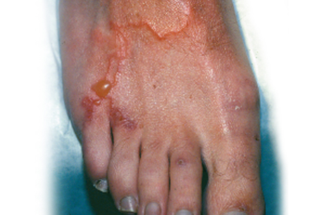 This may extend from a few millimeters to a few centimeters daily.[29]Feldmeier H, Jackson A, Heukelbach J, et al. A study in a community in Brazil in which cutaneous larva migrans is endemic. Clin Infect Dis. 2006 Jul 15;43(2):e13-8.
https://academic.oup.com/cid/article/43/2/e13/335778
http://www.ncbi.nlm.nih.gov/pubmed/16779735?tool=bestpractice.com
Associated pruritus is a key clinical finding and may be intense and uncomfortable, even preventing sleep.[2]Centers for Disease Control and Prevention. CDC Yellow Book 2024: health Information for international travel. Section 5: travel-associated infections & diseases - cutaneous larva migrans. May 2023 [internet publication].
https://wwwnc.cdc.gov/travel/yellowbook/2024/infections-diseases/cutaneous-larva-migrans
This may extend from a few millimeters to a few centimeters daily.[29]Feldmeier H, Jackson A, Heukelbach J, et al. A study in a community in Brazil in which cutaneous larva migrans is endemic. Clin Infect Dis. 2006 Jul 15;43(2):e13-8.
https://academic.oup.com/cid/article/43/2/e13/335778
http://www.ncbi.nlm.nih.gov/pubmed/16779735?tool=bestpractice.com
Associated pruritus is a key clinical finding and may be intense and uncomfortable, even preventing sleep.[2]Centers for Disease Control and Prevention. CDC Yellow Book 2024: health Information for international travel. Section 5: travel-associated infections & diseases - cutaneous larva migrans. May 2023 [internet publication].
https://wwwnc.cdc.gov/travel/yellowbook/2024/infections-diseases/cutaneous-larva-migrans
Vesiculobullous or papular lesions have been found to occur along the larval tracks in 10% to 40% of cases; some reports have noted bullae several centimeters in diameter.[3]Tremblay A, MacLean JD, Gyorkos T, et al. Outbreak of cutaneous larva migrans in a group of travellers. Trop Med Int Health. 2000 May;5(5):330-34.
http://onlinelibrary.wiley.com/doi/10.1046/j.1365-3156.2000.00557.x/full
http://www.ncbi.nlm.nih.gov/pubmed/10886795?tool=bestpractice.com
[30]Veraldi S, Arancio L. Giant bullous cutaneous larva migrans. Clin Exp Dermatol. 2006 Jul;31(4):613-4.
http://www.ncbi.nlm.nih.gov/pubmed/16716185?tool=bestpractice.com
Rarely, a returning traveler may present with folliculitis due to creeping larvae becoming trapped in the sebaceous follicular canal. In such cases, pruritic papules and pustules are found in association with relatively short tracks, primarily on the buttocks.[5]Caumes E, Ly F, Bricaire F. Cutaneous larva migrans with folliculitis: report of seven cases and review of the literature. Br J Dermatol. 2002 Feb;146(2):314-6.
http://www.ncbi.nlm.nih.gov/pubmed/11903247?tool=bestpractice.com
[31]Veraldi S, Persico MC, Francia C, et al. Follicular cutaneous larva migrans: a report of three cases and review of the literature. Int J Dermatol. 2013 Mar;52(3):327-30.
http://www.ncbi.nlm.nih.gov/pubmed/23414157?tool=bestpractice.com
Larval tracks may be single or multiple, and are located most commonly on the feet, thighs, and buttocks, related to the most common areas to come into contact with contaminated soil.[4]Davies HD, Sakuls P, Keystone JS. Creeping eruption. A review of clinical presentation and management of 60 cases presenting to a tropical disease unit. Arch Dermatol. 1993 May;129(5):588-91.
http://www.ncbi.nlm.nih.gov/pubmed/8481019?tool=bestpractice.com
[6]Jelinek T, Maiwald H, Nothdurft HD, et al. Cutaneous larva migrans in travelers: synopsis of histories, symptoms, and treatment of 98 patients. Clin Infect Dis. 1994 Dec;19(6):1062-6.
http://www.ncbi.nlm.nih.gov/pubmed/7534125?tool=bestpractice.com
However, lesions can occur on any unprotected part of the body, including hands, arms, trunk, scalp, face, breasts, and genitals.[32]Sow D, Soro F, Javelle E, et al. Epidemiological profile of cutaneous larva migrans in travelers returning to France between 2003 and 2015. Travel Med Infect Dis. 2017 Jun 15 [Epub ahead of print].
http://www.ncbi.nlm.nih.gov/pubmed/28624508?tool=bestpractice.com
Investigations
Diagnosis is based on clinical grounds and further investigation is rarely necessary.[2]Centers for Disease Control and Prevention. CDC Yellow Book 2024: health Information for international travel. Section 5: travel-associated infections & diseases - cutaneous larva migrans. May 2023 [internet publication].
https://wwwnc.cdc.gov/travel/yellowbook/2024/infections-diseases/cutaneous-larva-migrans
A minority of patients may demonstrate eosinophilia on complete blood count and/or elevated total IgE levels; however, these findings are nonspecific and these tests are therefore not recommended.[6]Jelinek T, Maiwald H, Nothdurft HD, et al. Cutaneous larva migrans in travelers: synopsis of histories, symptoms, and treatment of 98 patients. Clin Infect Dis. 1994 Dec;19(6):1062-6.
http://www.ncbi.nlm.nih.gov/pubmed/7534125?tool=bestpractice.com
[21]Blackwell V, Vega-Lopez F. Cutaneous larva migrans: clinical features and management of 44 cases presenting in the returning traveller. Br J Dermatol. 2001 Sep;145(3):434-7.
http://www.ncbi.nlm.nih.gov/pubmed/11531833?tool=bestpractice.com
[33]Shimogawara R, Hata N, Schuster A, et al. Hookworm-related cutaneous larva migrans in patients living in an endemic community in Brazil: immunological patterns before and after ivermectin treatment. Eur J Microbiol Immunol (Bp). 2013 Dec;3(4):258-66.
https://www.ncbi.nlm.nih.gov/pmc/articles/PMC3838541
http://www.ncbi.nlm.nih.gov/pubmed/24294495?tool=bestpractice.com
Skin biopsy or scrapings rarely identify migrating larvae because the larvae are usually located a few centimeters ahead of the creeping eruption, and skin biopsy should only be performed in cases of associated folliculitis.[5]Caumes E, Ly F, Bricaire F. Cutaneous larva migrans with folliculitis: report of seven cases and review of the literature. Br J Dermatol. 2002 Feb;146(2):314-6.
http://www.ncbi.nlm.nih.gov/pubmed/11903247?tool=bestpractice.com
[6]Jelinek T, Maiwald H, Nothdurft HD, et al. Cutaneous larva migrans in travelers: synopsis of histories, symptoms, and treatment of 98 patients. Clin Infect Dis. 1994 Dec;19(6):1062-6.
http://www.ncbi.nlm.nih.gov/pubmed/7534125?tool=bestpractice.com
There are no approved serologic or molecular diagnostic methods available. Epiluminescence microscopy is still an emerging technique. It is noninvasive and the larvae may be visualized migrating in the skin, although the sensitivity seems to be low.[34]Veraldi S, Schianchi R, Carrera C. Epiluminescence microscopy in cutaneous larva migrans. Acta Derm Venereol. 2000 May;80(3):233.
http://www.ncbi.nlm.nih.gov/pubmed/10954233?tool=bestpractice.com