Images and videos
Images


Evaluation of splenomegaly
Peripheral blood film of a patient with acute myeloid leukemia showing myeloid blasts with an Auer rod
From the collection Dr Priyanka Mehta; used with permission
See this image in context in the following section/s:
Evaluation of splenomegaly
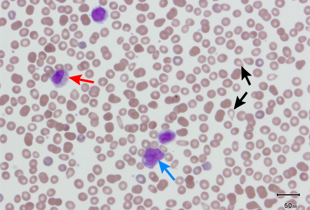
Peripheral blood smear showing leukoerythroblastic reaction: teardrop red blood cells (black arrows), and myelocyte (red arrow) and promyelocyte (blue arrow)
From the collection of Dr Ashkan Emadi and Dr Jerry L. Spivak; used with permission
See this image in context in the following section/s:
Evaluation of splenomegaly
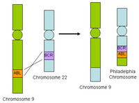
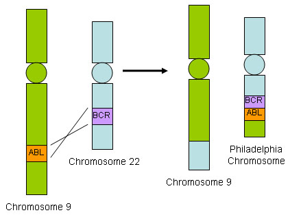
BCR-ABL translocation
From the collection of Dr Han Myint; used with permission
See this image in context in the following section/s:
Evaluation of splenomegaly
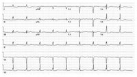
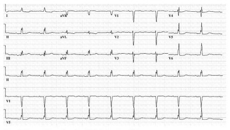
ECG of a patient with infective endocarditis. Note the first-degree AV block, nonspecific intraventricular conduction delay, and nonspecific ST-T wave abnormalities
From the collection of the Mayo Clinic Rochester, MN; used with permission
See this image in context in the following section/s:
Evaluation of splenomegaly
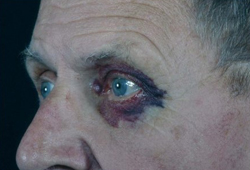
Classic periorbital purpura
From the collection of Dr Morie A. Gertz; used with permission
See this image in context in the following section/s:

Evaluation of splenomegaly
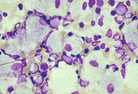
Bone marrow aspirate showing a typical Gaucher cell
From the collection of Dr Atul B. Mehta; used with permission
See this image in context in the following section/s:

Evaluation of splenomegaly
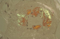
Congo red stain blood vessel in a bone marrow biopsy demonstrating green birefringence pathognomonic of amyloidosis
From the collection of Dr Morie A. Gertz; used with permission
See this image in context in the following section/s:
Evaluation of splenomegaly
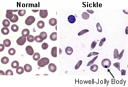
Red cells in sickle cell disease
From the collection of Dr Sophie Lanzkron; used with permission
See this image in context in the following section/s:
Use of this content is subject to our disclaimer