History and exam
Key diagnostic factors
common
pruritus
Virtually all patients with atopic dermatitis describe pruritus.
xerosis (dry skin)
Xerosis is a hallmark of atopic dermatitis.
sites of skin involvement
Infants typically show involvement of the cheeks, forehead, scalp, and extensor surfaces. Affected skin is often edematous, with prominent weeping and crusting. [Figure caption and citation for the preceding image starts]: Acute atopic dermatitis on the face of an infantFrom the personal collection of A. Hebert, MD; used with permission [Citation ends].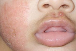
Children typically have involvement of flexures, particularly the wrists, ankles, and antecubital and popliteal fossae.[8][20]
In addition to the areas affected by acute disease, chronic atopic dermatitis often affects the neck, upper back, and arms, as well as the hands and feet.[1][5][64][Figure caption and citation for the preceding image starts]: Lichenification of the popliteal fossa in a child with atopic dermatitisFrom the personal collection of A. Hebert, MD; used with permission [Citation ends]. [Figure caption and citation for the preceding image starts]: Chronic dermatitis affecting the palm of a 64-year-old manFrom the personal collection of A. Hebert, MD; used with permission [Citation ends].
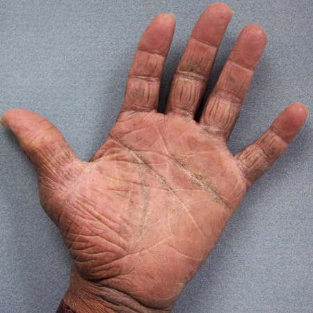
Overlap between acute and chronic atopic dermatitis may be seen due to the recurrent nature of the disease.[48]
Nuances in the visual appearance of atopic dermatitis can occur due to differences in pigmentation and distribution of lesions.[46] More well-demarcated lesions and increased scaling and lichenification are more common in Asian patients. Patients of African descent are less likely to develop flexural dermatitis, but rather present with extensor involvement as a more prominent feature.[46] Discoid or follicular patterns may be more common in Asian, black Caribbean, and black African children.[45]
Other diagnostic factors
common
erythema
Often noted in the acute flares.[48]
scaling
Often noted in the acute flares.[48]
vesicles
More common in acute flares and in infants.[48]
papules
More common in acute flares and in infants.[48]
keratosis pilaris
Follicular hyperkeratotic papules may be present on the extensor surfaces of the upper arms, buttocks, and anterior thighs, and are typically asymptomatic.[48]
excoriations
Commonly seen in areas that are easy to reach.[48]
lichenification
Thick, lichenified skin is evidence of chronic dermatitis.[48][Figure caption and citation for the preceding image starts]: Lichenification of the popliteal fossa in a child with atopic dermatitisFrom the personal collection of A. Hebert, MD; used with permission [Citation ends].
hypopigmentation
Seen in affected areas.[48][Figure caption and citation for the preceding image starts]: Hypopigmentation on the dorsal aspect of the hand in a 12-year-old girl with atopic dermatitisFrom the personal collection of A. Hebert, MD; used with permission [Citation ends].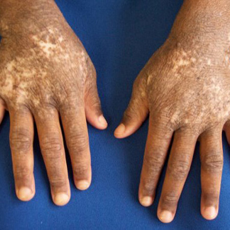 [Figure caption and citation for the preceding image starts]: Papules, lichenification, and hypopigmentation in a child with chronic atopic dermatitisFrom the personal collection of A. Hebert, MD; used with permission [Citation ends].
Risk factors
strong
filaggrin gene mutation
age <5 years
Usually presents in childhood; 45% of patients with atopic dermatitis are diagnosed by 6 months of age, and 70% to 85% of patients are diagnosed by the age of 5 years.[1][4][8]
Early onset of atopic dermatitis has been found to correlate with persistence of disease and more severe atopic disease within the atopic march.[34]
family history of atopic dermatitis
allergic rhinitis
weak
active and passive exposure to smoke
One systematic review and meta-analysis of observational studies concluded that active and passive exposure to smoke are associated with increased atopic dermatitis prevalence.[38]
Specifically, diagnosis of atopic dermatitis was associated with higher odds of exposure to passive smoke (OR 1.18, 95% CI 1.01 to 1.38) and active smoking (OR 1.87, 95% CI 1.32 to 2.63), but not to maternal smoking during pregnancy (OR 1.06, 95% CI 0.80 to 1.40).[38]
female sex
Atopic dermatitis affects females more than males in both the 6-7 years and the 13-14 years of age populations.[10]
African-American ethnicity
In the US, reported atopic dermatitis prevalence is 19.3% in African-American children and 16.1% in European-American children.[14]
Use of this content is subject to our disclaimer