Images and videos
Images

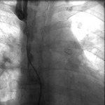


Superior vena cava syndrome
Venography showing superior vena cava stenosis. Stent placement in the left pulmonary artery is seen
Image obtained from cardiac catheterization laboratory at University of Missouri, Columbia; used with permission
See this image in context in the following section/s:
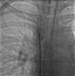
Superior vena cava syndrome
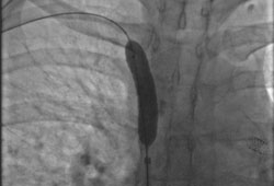
Percutaneous balloon angioplasty of the stenotic lesion in superior vena cava
Image obtained from cardiac catheterization laboratory at University of Missouri, Columbia; used with permission
See this image in context in the following section/s:

Superior vena cava syndrome
Supra- and infra-azygos obstruction leading to superior vena cava (SVC) syndrome. IVC: inferior vena cava
Reproduced with permission from Braunwald's Heart Disease, 8th ed (2008)
See this image in context in the following section/s:

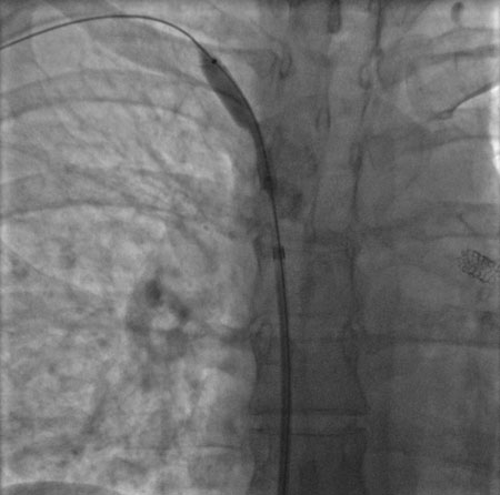
Superior vena cava syndrome
Stent deployment in the superior vena cava
Image obtained from cardiac catheterization laboratory at University of Missouri, Columbia; used with permission
See this image in context in the following section/s:
Superior vena cava syndrome
Postdilatation of the superior vena cava stent
Image obtained from cardiac catheterization laboratory at University of Missouri, Columbia; used with permission
See this image in context in the following section/s:
Use of this content is subject to our disclaimer