Approach
The diagnostic approach to a patient with tachycardia focuses on rapid assessment of the clinical consequences and careful evaluation to identify the mechanism of the arrhythmia and the setting in which it is occurring (drug toxicity, structural heart disease, ischemia).
In the setting of hemodynamic instability, it is important that diagnostic maneuvers do not delay therapy necessary to terminate the tachycardia.[1][2][50]
History
Often, patients with paroxysmal tachycardia will be asymptomatic at the time of evaluation. Symptoms associated with tachycardia include palpitations, fatigue, lightheadedness, presyncope, chest discomfort, and dyspnea.
Details of the pattern of the symptoms should be obtained, such as regular or irregular palpitations, number and frequency of episodes, potential triggers, whether the onset is abrupt or gradual, duration of symptoms, and how the tachycardia terminates.
The history should also include an evaluation of potential stressors such as hypovolemia, infection, ischemia, heart failure; the patient's medication regimen (including herbal supplements); a thorough history of substance use or misuse (including caffeine, energy drinks, and other stimulants); and any family history of arrhythmias or sudden death.
Presyncope and syncope associated with tachyarrhythmia and structural heart disease have a poor prognosis and should prompt detailed questioning surrounding the event.
A history of gradual onset and termination is more common with sinus tachycardia and atrial tachycardia, whereas an abrupt onset and termination is more common for reentrant tachycardias such as supraventricular tachycardia (SVT) and ventricular tachycardia (VT). The ability to terminate the rhythm abruptly with vagal maneuvers suggests that the reentrant rhythm circuit involves the AV node and is associated with AV nodal reentrant tachycardia (AVNRT) or orthodromic AV reciprocating tachycardia (AVRT).[14]
It is important to note that a common misconception is that hemodynamic stability may help to “rule out” VT. However, VT may commonly be tolerated hemodynamically and, in contrast, some atrial arrhythmias (for example, atrial fibrillation and atrial flutter conducting with a rapid ventricular rate) may be poorly tolerated. Therefore, hemodynamic stability should not be a factor in distinguishing VT from atrial arrhythmias.
Physical exam
The standard physical exam is often unrevealing, particularly if the tachycardia is episodic and not ongoing at the time of evaluation.
The physical exam should include a detailed cardiac examination to evaluate for valvular, congenital, and other structural heart disease.
A careful evaluation for signs of cardiomyopathy should be performed because of the worse prognosis of tachyarrhythmias associated with structural heart disease. This includes looking for S3 gallop, right ventricular (RV) heave, laterally displaced point of maximal impulse, and other signs of heart failure (elevated jugular venous pressure, lower-extremity edema).
If the patient has the symptoms at the time of physical evaluation, determining whether the pulse is regular or irregular can greatly assist with a diagnosis. Physical exam findings associated with AV dissociation such as cannon A waves or variability in the intensity of S1 are highly suggestive of VT.[51]
Diagnostic studies: 12-lead ECG
The resting 12-lead ECG is the cornerstone of the standard evaluation of tachycardia.
Even if the patient does not have the tachyarrhythmia at the time of the ECG, one can evaluate for evidence of prior myocardial infarction (pathologic Q waves), prolonged QT interval, ischemia, atrial or ventricular enlargement or hypertrophy, and signs of preexcitation.
Evidence of preexcitation, evidenced by a widened QRS complex with a delta wave, should raise suspicion for AVRT. The ECG should also be closely evaluated to rule out artifacts such as electrical interference or movement of telemetry monitoring leads as a cause for the observed abnormality. [Figure caption and citation for the preceding image starts]: Artifact overlying sinus rhythmFrom the collection of Robert W. Rho, MD; used with permission [Citation ends].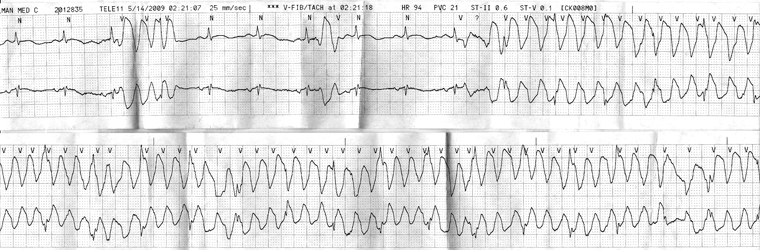
If the patient is experiencing the tachyarrhythmia at the time of the ECG, the findings can be diagnostic.
Having determined whether the tachyarrhythmia has a narrow or wide QRS interval, one can apply the appropriate diagnostic algorithm to develop a preliminary differential or diagnosis.[1] If P waves cannot be seen, sometimes an esophageal lead can also be used to help determine the atrial:ventricular relationship during tachycardia.[1][2]
During the evaluation of a regular narrow-QRS-complex tachycardia (<120 ms), assessment of the response to increased AV nodal blockade, either through carotid massage or with adenosine, can assist with the differential diagnosis. Intravenous adenosine should be administered in a monitored setting and with a continuous 12-lead ECG recording at the time of the maneuver. Absence of effect on ventricular rate, a temporarily gradual slowing of ventricular rate, or sudden termination of the tachycardia may be helpful in the diagnosis and can be therapeutic in some situations. Temporary AV block may unmask an atrial tachyarrhythmia. If this is ineffective or not feasible and the patient is hemodynamically stable, intravenous beta blocker or intravenous diltiazem or verapamil can be used.
For the evaluation of wide-QRS-complex tachycardia (>120 ms), differentiation between SVT and VT is important because intravenous medications for SVT (verapamil or diltiazem) can have adverse and even fatal consequences in a patient with VT. If differentiation between SVT and VT cannot be made, one should suspect and treat as VT until proven otherwise, especially in a patient with structural heart disease.
A useful way to subcategorize SVTs is to assess the relationship between the P wave and the preceding R wave. SVTs can be categorized into short-RP tachycardias (RP less than half the R-R interval) and long-RP tachycardias (RP greater than half the R-R interval). Short-RP tachycardia with retrograde P waves usually represents typical (slow-fast) AVNRT, AVRT, or atrial tachycardia with prolonged AV conduction. A long-RP tachycardia usually represents permanent junctional reciprocating tachycardia, atypical (fast-slow) AVNRT, or an atrial tachycardia conducting with brisk AV node conduction.[1][2]
Further diagnostic studies
Monitoring devices
If the patient has frequent (several per day) episodes of the presumed tachyarrhythmia but is not experiencing the rhythm at the time of evaluation, an ambulatory 24- or 48-hour Holter recording can be used.
If the episodes are less frequent and would be unlikely to occur during a 24- to 48-hour monitoring period, an event or wearable loop recorder can be used.[52][53]
If episodes occur rarely (<2 episodes per month) and are associated with hemodynamic instability or syncope (in which case activation of an event monitor is unlikely), an implantable loop recorder is appropriate and has been shown to be effective.[50]
If the patient already has a pacemaker or defibrillator in place, interrogation of the device may greatly assist with diagnosis.[5]
Direct to consumer technologies
Wearable and remote ECG-recording devices may be used by patients outside of a medical setting to provide single/multi-lead ECGs.[54][55] Some devices may not be approved as medical devices.
Clinicians should be prepared to discuss results generated using these technologies, and understand their risks and limitations.[56]
The 12-lead ECG remains the gold standard for the detection and evaluation of tachycardia.
Imaging
Imaging is not required in all cases and its use depends on the nature of the suspected tachyarrhythmia and the patient's overall clinical presentation.
Indications for echocardiography include wide-complex tachycardia of unknown origin, documented sustained atrial arrhythmias (atrial fibrillation, atrial flutter, SVT), or findings on history or physical exam that suggest structural heart disease.[57]
Exercise testing
Exercise testing may be helpful in defining the association of the arrhythmia to exercise, in provoking the arrhythmia, and in ruling out significant coronary artery disease, if appropriate.
If the arrhythmia is provoked during the study, the onset, termination, and a full 12-lead ECG of the arrhythmia is provided.
The exercise stress test is particularly helpful in provoking VT in patients suspected to have right ventricular outflow tract VT.
Electrophysiology testing
A diagnostic electrophysiologic study (EPS) is a useful tool for clarifying the mechanism of sustained and nonsustained supraventricular and ventricular arrhythmias. In wide-complex tachycardias where the diagnosis is uncertain, a diagnostic EPS can be helpful in establishing whether the arrhythmia is supraventricular or ventricular in origin. In addition to the diagnosis of tachyarrhythmias, several arrhythmias can be successfully treated with radiofrequency ablation at the time of the EPS.
Indications for referral to a cardiac arrhythmia specialist for EPS include, but are not limited to:
Unknown wide-complex tachycardias
Wolff-Parkinson-White syndrome (preexcitation with arrhythmia)
History of MI with history or symptoms suggestive of VT.
Laboratory testing
Baseline laboratory evaluation should be targeted toward the patient's overall clinical picture. But a number of tests should be included:
Blood electrolytes: particularly potassium, magnesium, and calcium; hypovolemia, which can manifest as prerenal azotemia or orthostatic hypotension, can cause sinus tachycardia
CBC: to determine if anemia is a contributing factor
Thyroid function tests: particularly if hyperthyroidism is in the differential diagnosis; results are often normal
Cardiac biomarkers: for patients who present with chest pain, have significant risk factors for ischemic heart disease, or are otherwise unstable; can show whether ischemia or infarction are contributing factors.
Drug levels: drug toxicity should be considered, particularly for patients who are on digitalis or who present with closely associated rhythms such as bi-directional VT or atrial tachycardia with AV block
Toxicology screen: for stimulants such as cocaine or tricyclic antidepressants.
Use of this content is subject to our disclaimer