Etiology
The different etiologies of portal hypertension are usually classified according to the site of obstruction to hepatic flow. Normal hepatic venous pressure gradient (HVPG) is 5 mmHg or less. Presinusoidal hypertension occurs when the HVPG is normal or less than the portal pressure. Intrahepatic/sinusoidal hypertension occurs when the portal free pressure equals the wedged hepatic venous pressure. Postsinusoidal hypertension occurs when the site of obstruction is distal to the sinusoids. Increased resistance to hepatic flow and/or increased hepatic flow together lead to the development of porto-systemic collaterals. These include those in the lower esophagus. Dilation and distention of these vessels occurs due to continued portal hypertension and results in variceal formation. The classification of portal hypertension according to the two major mechanisms is:
Increased resistance
Prehepatic:
Portal vein occlusion
Splenic vein occlusion
Congenital stenosis of the portal vein
Extrinsic compression of the portal vein.
Hepatic:
Presinusoidal
Sarcoidosis
Schistosomiasis
Congenital hepatic fibrosis
Primary biliary cholangitis
Idiopathic portal hypertension
Sinusoidal
Cirrhosis (all etiologies)
Alcoholic hepatitis
Postsinusoidal
Veno-occlusive disease
Budd-Chiari syndrome.
Posthepatic:
Constrictive pericarditis
Restrictive cardiomyopathy
Valvular heart disease
Web lesion of inferior vena cava.
Increased flow
Portal vein:
Myeloproliferative disorders.
Hepatic artery:
Hepatic artery-portal vein fistula.
Pathophysiology
The initial factor in the pathophysiology of portal hypertension is the increase in vascular resistance to portal blood flow. This results both from the structural distortion caused by the underlying disease (the mechanical component) and from active contraction of portal/septal myofibroblasts, activated hepatic stellate cells, and vascular smooth muscle cells in portal venules (the dynamic component).[20] Liver damage occurring in sepsis may itself contribute to an acute increase in portal hypertension.[21]
This active component of intrahepatic vasoconstriction accounts for 20% to 30% of the resistance.
Portal hypertension leads to the development of porto-systemic collaterals, possibly under the influence of angiogenic factors such as vascular endothelial growth factor (VEGF), allowing for shunting of blood around the liver. Portal hypertension persists despite the formation of collaterals because of splanchnic arteriolar vasodilation and insufficient decompression through the collaterals that have higher resistance than the liver. Clinically, gastroesophageal varices, together with ascites, are the most important consequence of portal hypertension.
Classification
Endoscopic appearance and liver function
In most centers using esophago-gastro-duodenoscopy (EGD), esophageal varices are graded according to their size, as follows:[6]
Small (Grade 1): small straight varices
Medium (Grade 2): enlarged tortuous varices occupying less than one third of the lumen
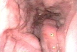
Large (Grade 3): large coil-shaped varices occupying more than one third of the lumen.[Figure caption and citation for the preceding image starts]: Large esophageal varicesFrom the personal collection of Gennaro d'Amico, MD [Citation ends].
 [Figure caption and citation for the preceding image starts]: Large esophageal varicesFrom the personal collection of Gennaro d'Amico, MD [Citation ends].
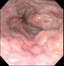
One consensus workshop recommended a simplified two-grade system (small and large) in 1990, with large varices being of a diameter >5 mm.[7] Recommendations for large varices apply to medium varices for centers using the three-grade classification.
Development of varices and their progression from small to large occur at a rate of approximately 7% to 8% per year.[4][8] The main factors associated with the development of varices, and their progression from small to large, are a hepatic vein pressure gradient (HVPG) >10 mmHg, decompensated cirrhosis (Child-Pugh B/C), alcoholic cirrhosis, and presence of red wale marks (defined as longitudinal dilated venules resembling whip marks on the variceal surface) at the time of baseline endoscopy.[5][8][9]
The most important predictors of hemorrhage are the size of the varices, the Child-Pugh class, and the endoscopic finding of red wale marks.[5]
Clinical classification of patients with cirrhosis at risk of esophageal varices and variceal bleeding[5]
Cirrhosis is classified into two prognostic stages: compensated and decompensated.
Decompensated cirrhosis is defined by ascites, variceal hemorrhage, or hepatic encephalopathy.
The compensated stage has been divided into further substages:
Patients with mild portal hypertension (hepatic vein pressure gradient [HVPG] >5 but <10 mmHg)
Patients with clinically significant portal hypertension (HVPG ≥10 mmHg):
without varices
with varices.
The following patient groupings provide a framework for both treatment and surveillance endoscopy:
Compensated cirrhosis with mild portal hypertension
Compensated cirrhosis with clinically significant portal hypertension, without gastroesophageal varices
Compensated cirrhosis with clinically significant portal hypertension, with gastroesophageal varices
Acute gastroesophageal variceal hemorrhage
Prevention of recurrent gastroesophageal hemorrhage.
North Italian endoscopic club for the study and treatment of esophageal varices[3]
Classification based upon size, severity of red wale marks, and Child-Pugh class:
Size of varices
Small
Medium
Large
Red wale markings
Absent
Mild
Moderate
Severe
Child-Pugh class
A
B
C
A risk stratification for variceal bleeding accompanies this classification, with cumulative scores for individual features added to define a risk class.
Size of varices
Small (<25% lumen radius) 8.7
Medium (25% to 50% lumen radius) 13.0
Large (>50% of lumen radius) 17.4
Red wale markings
Absent 3.2
Mild 6.4
Moderate 9.6
Severe 12.8
Child-Pugh class
A 6.5
B 13.0
C 19.5
Risk class according to score (the risk increases with increasing scores)
1 (<20)
2 (20 to 25)
3 (25.1 to 30)
4 (30.1 to 35)
5 (35.1 to 40)
6 (>40)
Use of this content is subject to our disclaimer