Tests
1st tests to order
hepatic venous pressure gradient (HPVG)
Test
The preferred method of assessing the presence of clinically significant portal hypertension.[5] However, noninvasive or minimally invasive techniques to assess portal hypertension have been proposed and are well established.[32]
Result
HVPG >5 but <10 mmHg indicates compensated cirrhosis with mild portal hypertension; HVPG ≥10 mmHg indicates compensated cirrhosis with clinically significant portal hypertension (with or without varices)
complete blood count
Test
Patients with low mean corpuscular volume and low Hb may have variceal bleeding, or, more probably, chronic gastrointestinal occult bleeding.
Low platelet count is indicative of portal hypertension resulting from cirrhosis.
Macrocytosis is often evident in patients with chronic alcoholism.
Result
microcytic anemia and/or thrombocytopenia
coagulation profile (INR/prothrombin time)
Test
Helpful in determining the synthetic functional capacity of the liver. Elevated INR/prothrombin time (PT) indicates that patient may have cirrhosis of the liver or liver failure.
Result
normal or elevated
serum LFTs
Test
Measures the severity of liver disease. Aminotransferases and bilirubin may be elevated if the patient has jaundice. Albumin may be decreased if patient is in liver failure. Total bilirubin may be normal in patients with compensated cirrhosis, but as the cirrhosis progresses, serum levels generally rise.
Result
elevated transaminases (with aspartate aminotransferase/alanine aminotransferase ratio ≥1), alkaline phosphatase, and bilirubin
BUN and creatinine
Test
Hyponatremia due to volume overload or use of diuretics may be present in patients with cirrhosis with ascites. BUN can be elevated secondary to prerenal azotemia, acute renal insufficiency, chronic renal insufficiency, or hepatorenal syndrome in cirrhosis of the liver.
Isolated elevated BUN (without elevated creatinine) is sometimes found as a result of breakdown of blood in the stomach in cases of acute bleeding.
Result
hyponatremia, elevated BUN and creatinine
blood typing/cross-matching
Test
Patients with variceal hemorrhage or upper gastrointestinal bleeding from other causes can experience rapid clinical deterioration. Blood should be sent for typing and cross-matching in the event that transfusion/blood products become necessary.
Result
variable
hepatitis B surface antigen (HBsAg)
Test
May indicate hepatitis B infection as cause of cirrhosis.
Result
positive
anti-hepatitis C virus IgG (anti-HCV IgG)
Test
May indicate hepatitis C infection as cause of cirrhosis.
Result
positive
esophago-gastro-duodenoscopy (EGD)
Test
Considered the most accurate method to identify varices.[5][6][33][34]
Findings may include: small varices (minimally elevated veins above the esophageal mucosa); medium varices (tortuous veins occupying less than one third of the esophageal lumen); and large varices (occupying more than one third of the esophageal lumen).
[Figure caption and citation for the preceding image starts]: (A) Grade I esophageal varices. These collapse to inflation of the esophagus with air. (B) Grade II esophageal varices. These are varices between grades 1 and 3. (C) Grade III esophageal varices. These are large enough to occlude the lumenTripathi D et al. Gut 2015;64:1680-704; used with permission [Citation ends].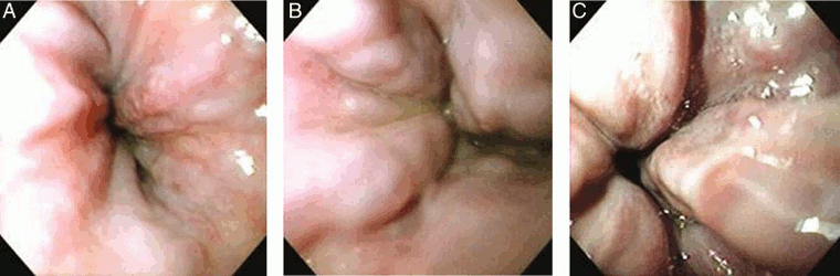
The most important predictor of hemorrhage is the size of varices, with the highest risk of first hemorrhage occurring in patients with large varices (15% per year).[3][4] The endoscopic finding of red wale marks (defined as longitudinal dilated venules resembling whip marks on the variceal surface) is also an important predictor of bleeding.[3][5]
Patients with a liver stiffness measurement (LSM) <20 kPa and platelet count >150,000/mm³ have a very low probability (<5%) of having high‐risk varices; therefore EGD can be safely avoided.[5]
Result
dilated veins in lower esophagus
liver stiffness measurement (LSM)
Test
Usually assessed by transient elastography; noninvasive test carried out by applying a vibrating probe to the thoracic wall at the level of the right liver lobe.[36] Velocity of wave propagation is directly proportional to liver stiffness.
Result
>25 kPa indicates clinically significant portal hypertension
Emerging tests
Use of this content is subject to our disclaimer