Images and videos
Images


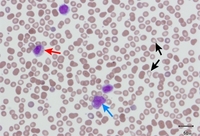
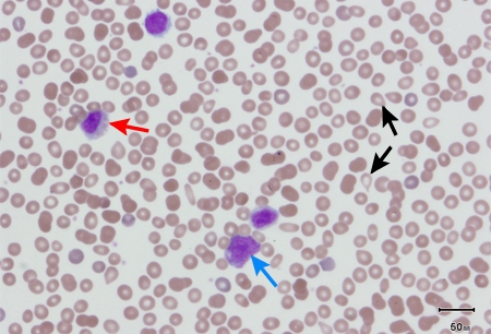
Evaluation of thrombocytopenia
Peripheral blood smear showing leukoerythroblastic reaction: teardrop RBCs (black arrows), and myelocyte (red arrow) and promyelocyte (blue arrow)
From the collection of A. Emadi and J.L. Spivak
See this image in context in the following section/s:
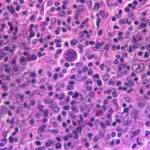
Evaluation of thrombocytopenia
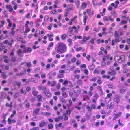
A diagnostic Reed-Sternberg cell is seen in the center of the image
From the collection of Dr C.R. Kelsey
See this image in context in the following section/s:
Evaluation of thrombocytopenia
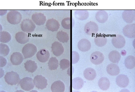
Thin-film Giemsa-stained micrographs showing ring-form Plasmodium vivax and P falciparum trophozoites
CDC Image Library/Steven Glenn, Laboratory & Consultation Division
See this image in context in the following section/s:
Evaluation of thrombocytopenia
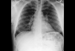
Bilateral hilar adenopathy, associated with sarcoidosis
From the collection of Muthiah P. Muthiah, MD, FCCP
See this image in context in the following section/s:
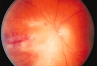
Evaluation of thrombocytopenia
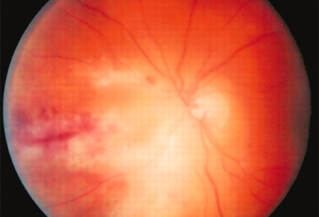
Fundoscopy (left eye) showing area of CMV retinitis inferonasally involving the vascular arcades and the optic disk, associated with vasculitis, and flame hemorrhages
Adapted from BMJ Case Reports 2009 (10.1136/bcr.02.2009.1576)
See this image in context in the following section/s:
Use of this content is subject to our disclaimer