Images and videos
Images


Osteosarcoma
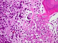
Osteoblastic osteosarcoma; lace-like osteoid in a highly pleomorphic sarcomatous stroma
Personal collections of Dr Michael J. Klein and Dr Luminita Rezeanu
See this image in context in the following section/s:
Osteosarcoma
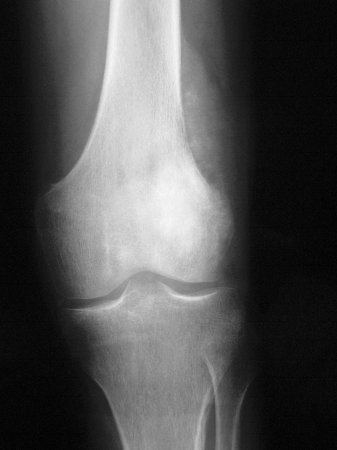
Conventional radiograph, anteroposterior view; poorly circumscribed, permeative lesion involving distal femoral metaphysis with mixed radiodense and radiolucent appearance; a large soft tissue mass with periosteal reaction is also present
Personal collections of Dr Michael J. Klein and Dr Luminita Rezeanu
See this image in context in the following section/s:

Osteosarcoma
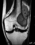
Magnetic resonance imaging, coronal view; osteosarcoma of distal femur showing low-intensity signal; T1-weighted image; actual intraosseous and extraosseous tumor extent is also appreciated
Personal collections of Dr Michael J. Klein and Dr Luminita Rezeanu
See this image in context in the following section/s:

Osteosarcoma
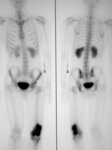
Bone scan; high radionuclide uptake at tumor site
Personal collections of Dr Michael J. Klein and Dr Luminita Rezeanu
See this image in context in the following section/s:
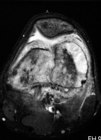
Osteosarcoma
Magnetic resonance imaging, axial view; osteosarcoma of distal femur showing high-intensity signal; T2-weighted image
Personal collections of Dr Michael J. Klein and Dr Luminita Rezeanu
See this image in context in the following section/s:
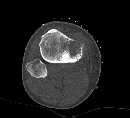
Osteosarcoma
Computed tomographic scan, axial view; osteosarcoma of proximal tibia; matrix production and bone destruction are best appreciated on conventional tomographs
Personal collections of Dr Michael J. Klein and Dr Luminita Rezeanu
See this image in context in the following section/s:
Use of this content is subject to our disclaimer