Approach
Differences in foot appearance and position noticed by parents most often reflect variations of normal physiologic development.[46] Unfamiliarity with normal lower-extremity growth and development, and desire for normal alignment in their children, may explain parents' concern and motivation to seek medical advice. Accurate diagnosis of torsional problems involves understanding the reason for consultation, taking parents' concerns seriously, taking a detailed history of the problem, performing a screening exam to rule out pathologic causes, and measuring severity with a rotational profile. Radiographic evaluation is rarely indicated unless a pathologic condition is suspected.
History
Evaluation begins with identifying the reason for consultation and having the parents describe the deformity and their concerns, expectations, and goals. Families may describe an angular deformity rather than a torsional problem. They may think that their child's abnormality may persist or cause long-term disability as they get older, or they may desire braces or special shoes to treat the problem. Practitioners should attempt to acknowledge the parents' concerns. Questions should be asked about the initial time of recognition of the problem, asymmetry, changes during that time interval, aggravating factors, any prior treatment, and family history of similar problems or inheritable conditions (e.g., vitamin D-resistant rickets, achondroplasia, mucopolysaccharidoses, or skeletal dysplasias). Out-toeing in a newborn is usually due to lateral rotation contractures at the hips, medial tibial torsion (MTT) usually presents in toddlers, and medial femoral torsion (MFT) usually presents at age 3 to 6 years. Parents should be asked about the child's sitting and sleeping habits. MTT is commonly associated with sitting on the feet, while MFT is associated with sitting in the W position. A complete maternal pregnancy, birth, and developmental history should be obtained. Standard developmental milestones (sitting, walking, talking, etc) should be determined. Infants usually sit by 8 months and walk independently by 12 to 18 months. A mature or adult walking pattern develops by 7 years. Pain of new onset or recent worsening, falling, tripping, delay in walking, tiring easily when walking, and limp may indicate an underlying disorder.
Miserable malalignment syndrome is typically seen in an older child with knee pain or instability. Infantile Blount disease is typically seen in a toddler >18 months old with varus angulation and medial tibial torsion. Obesity (>95% for the age) is typical. Early walking (before age 1 year) may be noted. The family history may be positive for Blount disease. In adolescent Blount disease, black males in the US are typically affected. Obesity is often present and medial knee pain may be noted.
Clubfoot is typically identified at birth. Skewfoot is typically identified at birth or may be iatrogenic following treatment of metatarsus adductus and clubfoot deformities.
Physical exam
Routine musculoskeletal screening exam should be performed before focusing on the rotational problem. A simple 18-step exam described by the Easter Seal Society should be performed on every child, even if parental concerns seem trivial.[47] This standard physical exam evaluates gait, coordination, balance, muscle tone, static deformities, and/or muscle contracture at each joint; torsional and other deformities of bone; and joint laxity. Tripping, falling, and shoe wear problems should be examined. Hip dysplasia or mild diplegia or hemiplegia related to cerebral palsy may present as torsional problems. Slipped capital femoral epiphysis should be considered in any older child or adolescent with complaints of knee, hip, or groin pain; gait abnormality; or asymmetric hip range of motion. Legg-Calve-Perthes disease may also be considered in any child with a limp, hip or groin pain, or asymmetric hip range of motion.
The key to establishing a diagnosis in torsional deformities is determining and recording a rotational profile, which uses anatomic landmarks around the knee, ankle, and foot.[1][2][48] The rotational profile includes measurements of:
Foot progression angle during gait [Figure caption and citation for the preceding image starts]: Foot progression angle (FPA) assessed while watching the child walk. FPA is formed by a line drawn in direction of walking and a line from the longitudinal axis of the foot. FPA is summation of torsional alignments of femur, tibia, and foot. Intoeing is designated as a negative number and out-toeing a positive numberFrom the collection of Lynn T. Staheli, MD [Citation ends].

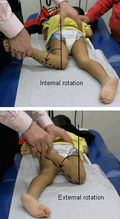
Arc of hip rotation (hip internal and external rotation) [Figure caption and citation for the preceding image starts]: Internal and external rotation of hip in extension is assessed with patient prone and knee flexed 90°From the collection of Tamir Bloom, MD [Citation ends].
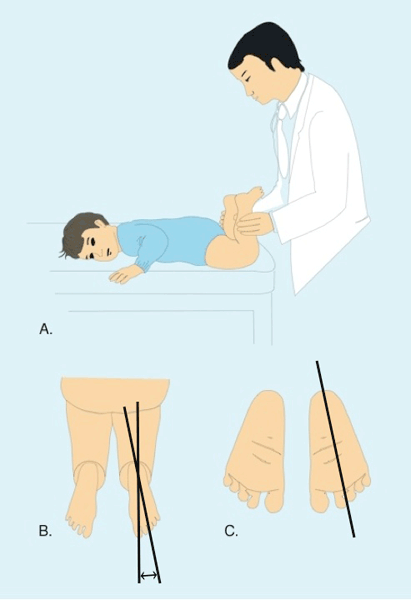
Thigh-foot angle [Figure caption and citation for the preceding image starts]: A,B: Thigh-foot axis assessed in prone position by measuring the angle between the longitudinal axis of the thigh and of the foot. C: Sole shape should be evaluated for forefoot adduction and abduction abnormalities such as metatarsus adductus. Heel-bisector line (drawn through midline axis of hindfoot and forefoot), in a normal foot, passes through the second web space. Lateral border of the foot is normally straightFrom the collection of Lynn T. Staheli, MD [Citation ends].
Transmalleolar axis: assesses the amount of tibial torsion present; this axis is the angle formed at the intersection of an imaginary line from the lateral to the medial femoral condyles, and a second line from the lateral to the medial malleolus
Heel-bisector line
Clinical values should be compared with published reference values to determine the relevance of findings based on physical exam. [Figure caption and citation for the preceding image starts]: Normal range and development of hip rotation throughout childhood. Green: normal ranges, mean ± 2 standard deviationsFrom the collection of Lynn T. Staheli, MD [Citation ends].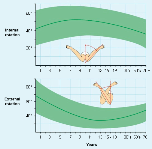 [Figure caption and citation for the preceding image starts]: Normal range and development of foot progression angle throughout childhood. Green: normal ranges, mean ± 2 standard deviationsFrom the collection of Lynn T. Staheli, MD [Citation ends].
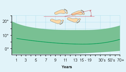 [Figure caption and citation for the preceding image starts]: Normal range and development of thigh-foot angle throughout childhood. Green: normal ranges, mean ± 2 standard deviationsFrom the collection of Lynn T. Staheli, MD [Citation ends].
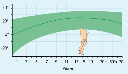
The anatomic definition of tibial torsion is not precise, and the optimal technique for clinical assessment is debatable. Tibial torsion is best assessed by the thigh-foot angle, the transmalleolar axis (TMA), and the second-toe test.[1][25][49] The TMA may be more accurate than the thigh-foot angle and second-toe test in children with foot deformities.[50] Another technique for measuring tibial torsion is the footprint method.[51] With the patient sitting, the footprint is traced on precisely placed lined paper. A line is then drawn connecting the 2 malleoli. The angles between the malleoli and any line on the paper are used to determine the TMA.
Femoral anteversion is evaluated by comparing internal and external rotation of the hip in the prone position, as well as palpating the point of maximal trochanteric prominence.[52][53][54] Prone measurements of hip rotation are more accurate than supine measurements because the pelvis is stabilized.
Angular deformities commonly coexist with rotational abnormalities and should be assessed by measuring the intercondylar or transmalleolar distance. These measurements, including the rotational profile, can then be compared with published reference values.[2][55]
Clinical exam in the office setting is limited, because it is primarily based on static evaluation of the lower extremity, whereas functional activities, such as walking, are dynamic.[56] In addition, torsional measurement may inaccurately assess deformity because of excessive joint laxity, joint contractures, and muscle spasticity, and when torsional deformities exist along with angular deformities.[2][57][58][59]
Neuromuscular conditions are determined through neurologic exam, including testing of primitive reflexes, postural and protective reactions, and motor tone. Abnormal movement patterns help to identify a child with a potential neuromuscular condition. Many conditions have multiple orthopedic components; the exam should not focus on one body part.
Miserable malalignment syndrome may demonstrate a medially pointing patella ("squinting patella"). A W sitting position may be present and the Q angle (formed from a line drawn from the anterior superior iliac spine to the center of the kneecap, and from the center of the kneecap to the tibial tubercle) may be increased. [Figure caption and citation for the preceding image starts]: Photo of a child sitting in the W positionFrom the collection of Tamir Bloom, MD [Citation ends].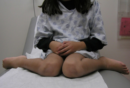
In slipped capital femoral epiphysis, out-toeing or asymmetric hip range of motion in an adolescent is a possible sign.
A convex lateral border indicates forefoot adductus seen in metatarsus adductus with heel-bisector line hitting lateral to second web space and normal ankle range of motion. It is more frequently seen on the left and in the first year of life.
Flat feet may be seen in out-toeing. They are normally painless and not tender on exam. Assessing flexibility demonstrates an arch when the foot is raised or when the first metatarsophalangeal joint is passively dorsiflexed. The hindfoot inverts with ankle plantarflexion (standing on the balls of the feet).
In infantile Blount disease the cover-up test may be positive.[60] This qualitatively assesses the alignment of the proximal portion of the shank or lower leg relative to the thigh or upper leg. Neutral or varus alignment is considered a positive test.[60] Lateral knee thrust may be present in both adolescent and infantile Blount disease.
In skewfoot, the hindfoot is in valgus, the midfoot is abducted, and the forefoot is adducted.
In clubfoot, the equinus contracture with limited dorsiflexion of the ankle may be noted.
Radiographic evaluation
Radiographic evaluation is not needed during initial evaluation of most torsional problems. Radiographic techniques are generally not superior to clinical assessment.[52] Do not order radiographs for a child <8 years with simple in-toeing gait, which can be monitored unless there is severe tripping and falling, or asymmetry.[46][61] In case of suspected hip pathology (e.g., slipped capital femoral, hip dysplasia) an anteroposterior pelvis and frog lateral views should be obtained. However, for a child <4 months old, radiographs are usually not appropriate when assessing for developmental dysplasia of the hip.[62] Cross-table lateral film should be ordered when a child >8 years has had a recent change in gait and knee or hip pain. Foot x-rays help in the diagnosis of clubfoot, skewfoot, congenital vertical talus, and hallux valgus.
With miserable malalignment syndrome, standard radiographic anteroposterior, lateral, and Merchant views are used to evaluate for patella alta, patellar subluxation or dislocation, and trochlea morphology.
In Blount disease, the medial epiphyseal line may demonstrate irregularity and abnormal sloping of the medial tibial epiphysis. The metaphyseal portion of the plate may demonstrate irregular beaking.[63][64] Imaging with standing anteroposterior x-ray of the lower extremities, including the hips and ankles, with the lower extremities rotated such that the knees (not the feet) are aligned straight ahead, is indicated in children >18 months of age with a positive cover-up test, asymmetric deformity, and family history of bowing or severe deformity.
With slipped capital femoral epiphysis, the epiphyseal plate is widened and the epiphysis appears somewhat shortened on the affected side and the margin of the metaphysis may appear indistinct on anteroposterior pelvis and lateral hip x-ray.
In skewfoot, standing anteroposterior and lateral x-rays of the foot confirm diagnosis. Z configuration of the foot on anteroposterior radiograph, with abduction of the midtarsal joints and adduction of the metatarsals, is seen. There is usually plantar flexion of the talus on the lateral x-ray.
Other investigations
Three-dimensional computed tomography (CT), magnetic resonance imaging (MRI), and ultrasound all accurately and precisely measure torsional alignment.[65][66][67] However, do not order CT or MRI in children until all appropriate clinical and plain radiographic exams have been completed.[61] CT ("gunsight" scan) evaluation of rotational profile may be used to measure severe or complex torsional deformities, such as miserable malalignment syndrome, in older children.[58][68][69] Concerns exist about excessive radiation exposure and measurement variability with plain radiographs and, even more so, with CT scan. If additional imaging is needed for preoperative evaluation, an MRI[70][71] or EOS™[72][73][74]low-dose biplanar x-ray may be preferable to CT scan. The availability, cost, and need for sedation (with MRI) should be considered when contemplating advanced imaging for torsional abnormalities in children.
[Figure caption and citation for the preceding image starts]: Severe torsional deformities may be assessed with gunsight CT scan by measuring the angle between the transverse axes on CT cuts of the proximal and distal juxta-articular regions© 1980 J Bone Joint Surg Br. Reprinted from: J Bone Joint Surg Br. 1980;62-B:238-242, with permission [Citation ends]. Quantitative gait analysis may provide information on dynamic alignment and range of motion. It is reserved for assessing gait abnormalities in neuromuscular conditions for surgical decision-making.
Use of this content is subject to our disclaimer