Approach
Uterine prolapse is diagnosed by clinical exam and staged according to the extent of downwards displacement of the most affected vaginal area.[24]
Clinical history
Key risk factors for prolapse include previous vaginal delivery, advancing age, obesity, previous surgery for prolapse, family history of prolapse, white ancestry, connective tissue disorders, and increased intra-abdominal pressure (e.g., chronic obstructive airway disease, chronic constipation with excessive straining, heavy lifting, and hard physical activity).
The most specific symptom of pelvic organ prolapse (POP) is the patient's report of seeing or feeling the vagina or cervix protrude through the vaginal opening, which also correlates with severity.[20] The patient frequently describes a sensation of bulging, fullness, or heaviness in the pelvic area. The heaviness is often described as a dragging discomfort or pressure in the lower abdomen or pelvis. In certain cases, voiding or storage dysfunction accompanies pelvic discomfort, presenting with urinary retention or urinary incontinence, respectively.[19] Women progressively become more symptomatic after prolonged standing or physical exertion. Defecatory dysfunction and sexual dysfunction (dyspareunia) may coexist. These concomitant disorders are probably not directly related to POP, although they share similar risk factors, and may persist after otherwise successful anatomic repair.[Figure caption and citation for the preceding image starts]: Apical prolapse POPQ stage IIIFrom the collection of Prof L. Brubaker and Dr L. Lowenstein; used with permission [Citation ends].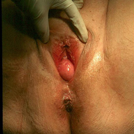 [Figure caption and citation for the preceding image starts]: Total uterovaginal prolapse (procidentia) POPQ stage IVFrom the collection of Prof L. Brubaker and Dr L. Lowenstein; used with permission [Citation ends].
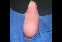
Physical exam
Any woman with gynecologic complaints related to POP symptoms should undergo pelvic exam.[25]
Physical examination should be performed with the patient resting and straining in both supine and standing positions.
Pelvic floor muscle tone should be assessed.
Complete gynecologic exam, including bimanual exam, is needed for estimating uterus size and cervical length.
Vaginal mass is palpated on digital exam.
A vaginal speculum is used to view the cervix and vaginal vault, while the patient is performing a valsalva maneuver. The fixed blade of the disassembled speculum is placed to support the posterior vaginal wall for evaluation of an apical or anterior prolapse. Whereas, the anterior vaginal wall is supported for evaluation of a posterior prolapse.[Figure caption and citation for the preceding image starts]: Apical prolapse POPQ stage IIIFrom the collection of Prof L. Brubaker and Dr L. Lowenstein; used with permission [Citation ends].[Figure caption and citation for the preceding image starts]: Total uterovaginal prolapse (procidentia) POPQ stage IVFrom the collection of Prof L. Brubaker and Dr L. Lowenstein; used with permission [Citation ends].
A systematic pelvic organ prolapse quantification (POP-Q) exam defines the extent of the prolapse and establishes the segments of the vagina that are affected (anterior, posterior, or apical).[22] See Criteria.
Investigations
Women with advanced (stage 3 or 4) prolapse often have voiding difficulties, which may result in urinary retention, recurrent urinary tract infection, and, in rare cases, damage to the renal parenchyma.[22] Lower urinary tract function should be assessed by evaluation of urine loss and type (stress or urgency urinary incontinence) and adequacy of bladder emptying.[22]
If the prolapse is beyond the hymen or the patient has concomitant incontinence, assessment of the postvoid residual urine volume is important, particularly if surgical intervention is planned. Postvoid residual urine volume may indicate voiding dysfunction, which may progress to complete retention following surgical intervention.[19][22][26] Before consenting patients for vaginal surgery to repair POP, an assessment for occult stress urinary incontinence is indicated, by performing a cough stress test or urodynamic testing after reduction of the prolapsed uterus by pessary or swab.[19] Urinalysis is also recommended as part of the initial assessment of patients with low urinary tract symptoms, as infection is known to exacerbate incontinence symptoms.[27][28][29]
Use of this content is subject to our disclaimer