Approach
The diagnosis of an anorectal abscess is usually suspected from a patient's clinical history and confirmed by physical examination. Laboratory and radiologic studies are not usually needed for the diagnosis of an anorectal abscess but can be useful in some special situations.
The location of an anorectal abscess affects its diagnosis and management.[7] Intersphincteric abscesses are difficult to diagnose because they produce little swelling and few perianal signs of infection, but they are associated with anal pain that is so severe that it precludes digital rectal examination. Anesthesia is usually required for an adequate examination and diagnosis of the condition.[16] Supralevator abscesses may present with symptoms that mimic an intra-abdominal condition. Rectal examination usually reveals a tender, indurated area above the anorectal ring, but imaging with computed tomography (CT) or magnetic resonance imaging (MRI) may be required to make the diagnosis.[10]
History
The presence of key risk factors such as a history of Crohn disease or anal fistula should be elicited. In addition, it should be noted that anorectal abscesses are more common in men than women.[4][14]
Patients with anorectal abscesses usually relate a history of localized anal or perianal pain.[2] Pain usually begins 1 to 2 days before presentation and becomes progressively more severe. Patients frequently complain of swelling and warmth of the perianal tissues. The patient may occasionally relate the onset to some precipitating event such as a difficult bowel movement, though pain associated only with defecation is likely to be due to a fissure. The pain may be exacerbated by movement, coughing, sneezing, or bowel movements. Patients may often try taking warm baths as pain relief, but these fail to improve their pain or make it worse. Most patients will not report rectal bleeding unless their abscess has spontaneously drained (usually associated with some decrease in the pain). Fever is common and is usually <101.5ºF (38.6ºC).
Patients with rare supralevator abscesses may describe pain in the lower abdomen or pelvis, mimicking an intra-abdominal condition.[10] Occasionally patients with anorectal abscesses may complain of being unable to urinate, particularly men with a previous history of difficulties with urination.[2][8] Symptoms of inflammation, pain, and swelling are frequently absent or diminished in patients with:[2]
Diabetes
Immunocompromise
Debilitation
Older age
Associated necrotizing soft-tissue infection.
Physical examination
An adequate anorectal examination can usually be performed in the office or emergency department, though on occasion this may be impossible because of pain. Intersphincteric and supralevator abscesses in particular require anesthesia for full examination.
The most common finding on physical examination is a tender, indurated area immediately adjacent to the anus, within the anal canal, or above the anorectal ring.[2] The further the indurated area is located from the anal verge, the less likely it is to be an anorectal abscess. Anal fistulae associated with anorectal abscesses may have a hard, cord-like structure leading toward the anus and palpable in the soft tissues. Infected epidermal inclusion cysts are much more likely when the indurated area is more than 3 cm from the anal verge while pilonidal disease is more common when the induration is located in the intergluteal area.
Occasionally, the infection can spread to involve both ischiorectal fossae and the postanal space (horseshoe abscess), though anorectal abscesses are almost always solitary. The induration in horseshoe abscesses may be more prominent in the ischiorectal fossae and appear to be bilateral abscesses.[17] If multiple abscesses are present, the diagnosis is much more likely to be multiple infected epidermoid inclusion cysts or perianal hidradenitis suppurativa.
Induration may be absent or diminished in older adults, patients with diabetes, or in those who are immunocompromised or debilitated. Detection of low-grade fever and mild tachycardia should also form part of the physical examination as these are common symptoms of anorectal abscess.
Laboratory studies
Laboratory studies are rarely helpful in the diagnosis and management of anorectal abscesses and need not be a routine component of the evaluation and management of these patients.[2]
Basic tests include the following.
WBC count: will frequently reveal a leukocytosis with an increased proportion of neutrophils. Anorectal abscesses do not cause anemia or other hematologic abnormalities.
Blood glucose: may show hyperglycemia, which may or may not be associated with diabetes.
For patients with a necrotizing soft-tissue infection related to their anorectal abscess, serum electrolyte determination may reveal an elevated BUN and creatinine, decreased bicarbonate, and an increased base deficit (metabolic acidosis).
Abnormal blood chemistry results should generally be evaluated further only after the acute abscess has been treated, while addressing any urgent treatment considerations acutely (e.g., volume depletion, hyperglycemia).
In the past, some experts recommended culture of the contents of an anorectal abscess, feeling that the bacteriology of the abscess could be predictive of the presence of an associated anal fistula.[18][19] Culture of the contents of an anorectal abscess is usually reserved for patients with recurrent abscesses without a fistula identified, or for those with risk factors for one of the rare causes of anorectal abscess such as HIV or immunosuppression, or patients from a developing region. Specific microbiologic and culture techniques may be needed to identify these unusual pathogens.[20][21]
Radiologic studies
Radiologic studies are rarely helpful in the diagnosis and management of anorectal abscesses. Occasionally, for patients with complex or atypical presentations, or those with supralevator or horseshoe abscesses, anal ultrasonography has been used for evaluation. However, the severe pain associated with the anorectal abscess frequently limits the use of this modality. Other imaging modalities such as CT or MRI may be more helpful in the evaluation of these patients.[4][5][6][Figure caption and citation for the preceding image starts]: CT demonstrating a perirectal abscessFrom the collection of Dr C. Neal Ellis; used with permission [Citation ends].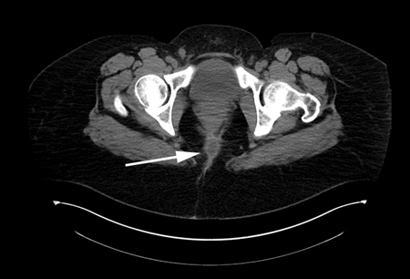
Use of this content is subject to our disclaimer