Etiology
Clubbing may be acquired or present as a hereditary anomaly.
Pulmonary
Lung cancer
Clubbing may be seen with any cell type of lung cancer.
Most frequently associated with squamous and adenocarcinomas and least frequently with small cell carcinoma.[11]
Bronchiectasis
Disease process characterized by abnormally dilated bronchi with thickened bronchial walls.
More common in middle-aged and older people; usually due to an infectious cause.
Underlying conditions associated with bronchiectasis include primary ciliary dyskinesia, Kartagener syndrome, cystic fibrosis, and diffuse panbronchiolitis.[12]
Lung abscess
Risk factors include immunosuppression, poor oral hygiene, drug misuse, alcohol misuse, seizure disorders, stroke, and lung cancer.
Empyema
Most commonly results from untreated bacterial infection in the pleural space.
Pulmonary metastases
Caused by a number of primary tumors (e.g., colorectal cancer, bone and soft-tissue sarcoma, melanoma, renal cell carcinoma, breast cancer, germ cell tumor).
Pulmonary tuberculosis
Pooled prevalence of digital clubbing of 17.1% reported in children and adolescents with tuberculosis.[13]
Clubbing is not usually a feature of pulmonary tuberculosis (TB) that occurs in isolation.
Can occur in cavitating TB and in patients who have pulmonary TB with underlying HIV infection.
Cystic fibrosis (CF)
Reported pooled prevalence of digital clubbing and hypertrophic osteoarthropathy of 19.5% and 5.0%, respectively, in children and adolescents with CF.[13]
CF is characterized by multiple organ system involvement, mainly resulting in chronic respiratory tract infections and the effects of pancreatic enzyme insufficiency.
Interstitial pulmonary fibrosis
A group of conditions involving chronic fibrosing interstitial lung disease of varying causes. May be idiopathic.
Pooled prevalence of digital clubbing of 31.3% reported in adults with interstitial lung diseases.[13]
Characterized by dyspnea, reduced lung volumes, and poor gas exchange on lung function testing.[14][15]
Digital clubbing has been associated with parameters of disease severity in patients with interstitial lung disease.[16]
Sarcoidosis
Chronic granulomatous disorder, characterized by accumulation of lymphocytes and macrophages in the lungs and other organs.
Lungs and intrathoracic lymph nodes are involved in >90% of patients, but virtually any organ can be affected.[17]
Asbestosis
Diffuse interstitial fibrosis of the lung as a consequence of chronic inhalation of asbestos fibers.[18]
Occupational history is important, as the type of work performed usually indicates asbestos exposure (shipyard, construction, maintenance, vehicle brakes, asbestos cement, and insulation or production of tiles, shingles, gaskets, or textiles).
May lead to a number of respiratory diseases, including lung cancer, pleural plaques, benign pleural effusions, and malignant mesotheliomas.
Risk of lung cancer increased in patients exposed to cigarette smoke.
Pleural mesothelioma
Classified into 3 types based on thoracoscopic biopsy and cytology samples: epithelial, sarcomatoid, and mixed.
About 80% of patients have had occupational exposure to asbestos.[19]
Lipoid pneumonia
Rare form of pneumonia that may be due to release of endogenous lipids in the lung (when lung tissue breaks down distal to an obstruction, e.g., lung carcinoma, bronchiolitis obliterans, or following lung necrosis after chemotherapy and radiation therapy) or caused by inhalation of exogenous lipids (e.g., aspiration of ingested oil, including mineral and vegetable oil).[20]
Pulmonary artery sarcoma
Rare sarcoma arising from the intima of the pulmonary artery.
Hypersensitivity pneumonitis (extrinsic allergic alveolitis)
Is the result of non-IgE mediated immunologic inflammation of the alveoli and distal bronchioles caused by repeated inhalation of nonhuman protein, such as from plant, bacteria, or animal, or the result of a chemical conjugated to a human airway protein, such as albumin.
Clubbing occurs in approximately half of those with chronic hypersensitivity pneumonitis.[21][22]
Cardiovascular
Congenital heart disease
Clubbing is a common sign of cyanotic congenital heart disease (e.g., tetralogy of Fallot, double outlet right ventricle, transposition of great vessels).
May also occur in association with other cardiac causes of right-to-left shunting such as patent ductus arteriosus, coarctation of the aorta, and in ventral septal defects.[23]
Infective endocarditis
Infection of the endocardial surface of the heart, including the valvular structures, chordae tendineae, site of septal defects, or the mural endocardium.[24][25]
May be acute, typically developing over a period of days to weeks, or subacute, typically developing over the course of weeks to months.
Pooled prevalence of digital clubbing of 27% reported in adults with infective endocarditis.[13]
Atrial myxoma
Most common of the rare benign primary cardiac tumors.
Usually found in the left atrium and typically attached to the septum.
Axillary artery aneurysm
Rare but potentially life-threatening cause of unilateral clubbing.
Majority are pseudoaneurysms that arise after trauma or injuries. True aneurysms seldom occur.
Brachial arteriovenous malformations
Abnormal connection between veins and arteries occurring in the arm.
Rare condition causing unilateral clubbing.
Usually congenital.
Hepatobiliary
Pooled prevalence of digital clubbing of 22.8% reported in adults with hepatic diseases.[13]
Primary biliary cholangitis
Autoimmune disease of the liver marked by slow and progressive destruction of bile canaliculi within the liver.
More common in women than men, and marked by intense itching.[26]
Cirrhosis of the liver
Pathologic end-stage of any chronic liver disease.
Most commonly results from chronic hepatitis B and C infection, alcohol misuse, and metabolic dysfunction-associated steatotic liver disease (previously known as nonalcoholic fatty liver disease).
Leads to portal hypertension, liver insufficiency, and hepatic failure.
Generally considered to be irreversible in its advanced stages.
Gastrointestinal
Pooled prevalence of digital clubbing of 33.4% reported in adults with intestinal disease.[13]
Ulcerative colitis
Form of inflammatory bowel disease that affects the rectum and extends proximally.
Characterized by diffuse inflammation of the colonic mucosa and a relapsing, remitting course.
Crohn disease
Inflammatory bowel disease that may involve the entire gastrointestinal tract.
Strictures, abdominal pain and fistulae are common.
Leiomyoma of the esophagus
Rare benign tumor arising from the smooth muscle of the esophageal submucosa, jutting into the esophageal lumen.
Achalasia
Esophageal motor disorder characterized by loss of peristalsis in the distal third and failure of the lower esophageal sphincter to relax in response to swallowing.
Ulcerative esophagitis
May be caused by alcohol, drugs, GERD, or peptic ulceration.
Celiac disease
Systemic autoimmune disease triggered by dietary gluten peptides found in wheat, rye, barley, and related grains.
Immune activation in the small intestine leading to villous atrophy, hypertrophy of the intestinal crypts, and increased numbers of lymphocytes in the epithelium and lamina propria.
Locally, causes gastrointestinal symptoms and malabsorption.
Diverse systemic manifestations, potentially affecting almost every organ system.
Tropical sprue
Acquired malabsorptive condition of probable infectious etiology.
Features include altered small-bowel mucosa, chronic diarrhea, and signs and symptoms of multiple vitamin and nutrient deficiencies.
Seen in both natives of, and long-term visitors to, specific endemic areas in the tropics.
Endocrine
Acromegaly
Rare chronic disease caused by excessive secretion of growth hormone, usually due to a pituitary somatotroph adenoma.
Severe secondary hyperparathyroidism
Most commonly caused by chronic renal insufficiency.
Characterized by excessive secretion of parathyroid hormone in response to hypocalcemia.
Thyroid acropachy
Extreme manifestation of Graves disease or autoimmune thyroiditis, causing soft-tissue swelling and digital clubbing.
Nonpulmonary malignant
Hodgkin lymphoma (Hodgkin disease)
Uncommon hematologic malignancy arising from mature B cells.
Characterized by orderly spread of tumor from one group of nodes to the other and the presence of Reed-Sternberg cells on histopathology.
Disseminated chronic myeloid leukemia
Malignant clonal disorder of hematopoietic stem cells.
Multiple organ system involvement regarded as disseminated disease.
Thyroid cancer
Most commonly presents as an asymptomatic thyroid nodule detected by palpation or ultrasound in a woman in her 30s or 40s.
Histologic variants include papillary, follicular, medullary, and anaplastic neoplasms.
Thymus cancer
Rare form of cancer.
Myasthenia gravis, lupus, and rheumatoid arthritis possible predisposing factors.
Hereditary
Familial clubbing
Clubbing without any associated signs or symptoms.
Exact genetics undefined; most likely inherited as an autosomal-dominant trait.[27]
Sometimes regarded as incomplete form of primary hypertrophic osteoarthropathy (HOA).[28][29][30]
Important to exclude other causes of clubbing before reaching this diagnosis.
Pachydermoperiostosis
Rare autosomal-dominant inherited condition.
Regarded as primary HOA and accounts for 5% of all cases of HOA.[31]
Characterized by the combination of pachydermia (elephant-like skin), periostosis (swelling around the joints, especially the wrists and knees), and clubbing.[32]
HOA may result from a gene mutation in SLCO2A1, which encodes the prostaglandin transport protein.[33][34][Figure caption and citation for the preceding image starts]: Pachydermoperiostosis: furrowed and thickened foreheadFrom the collection of Dr Murlidhar Rajagopalan [Citation ends].
 [Figure caption and citation for the preceding image starts]: Pachydermoperiostosis: finger clubbingFrom the collection of Dr Murlidhar Rajagopalan [Citation ends].
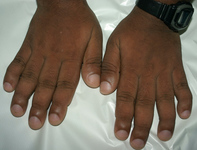 [Figure caption and citation for the preceding image starts]: Pachydermoperiostosis: wrist joint swellingFrom the collection of Dr Murlidhar Rajagopalan [Citation ends].
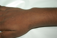
Other
Secondary hypertrophic osteoarthropathy (HOA)
Syndrome characterized by proliferative changes in the skin and skeleton. Rarely, may present as a presumptive acute reactive arthritis.[35]
Enlargement of the extremities due to periarticular and osseous proliferation as well as digital clubbing are common.
Associated with cardiopulmonary diseases and malignancies (e.g., lung cancer, lung abscess, bronchiectasis, emphysema, Hodgkin lymphoma, metastatic disease, cystic fibrosis, cirrhosis, inflammatory bowel disease, biliary atresia).
Pooled prevalence of HOA of 10.1% in adults with cancers.[13]
Presence of an underlying condition differentiates HOA from pachydermoperiostosis (considered primary HOA), which does not require immediate attention or intervention.[36]
Palmoplantar keratoderma
May be inherited (either autosomal dominant or autosomal recessive) or acquired (due to changes in an individual's health or environment).
Characterized by an abnormal thickening of the palms and soles.
Fischer syndrome and Volavsek syndrome (also called Buschke-Fischer-Brauer syndrome) are considered variants of palmoplantar keratoderma.[37]
Pregnancy
In rare cases, increased vascular demand of peripheries and hormonal factors can induce clubbing in pregnancy.
Pseudoclubbing
Lovibond angle (the angle made by the proximal nail fold and nail plate) between 160° and 180° possibly reflects early stages of clubbing or a pseudoclubbing phenomenon.
Unlike digital clubbing, there is no clear definition of pseudoclubbing.[38] In addition, there is no convincing etiopathogenic mechanism for pseudoclubbing. Asymmetric finger involvement has been observed in the majority of cases. Acro-osteolysis is also seen, but is not a discriminatory feature as it also occurs in true clubbing.[38]
Myelofibrosis
Reactive process common to many malignant and benign disorders.
Clubbing has been described in myelofibrosis.[39]
Use of this content is subject to our disclaimer