Approach
Peripheral edema is a physical examination finding. As such, the only diagnostic test to establish the presence of edema is examination and palpation of the extremities. Diagnosing the underlying cause of the edema is the goal of the investigative evaluation.
History
Patients report swelling and a tense sensation in the extremities, usually worse the longer the extremity remains dependent (subject to the influence of gravity). Edema may accumulate around the thighs, buttocks and sacrum in nonambulatory patients.
The affected limb often feels heavy or weak.
Patients may describe feeling as though clothing or jewelry no longer fit, or may indicate changes in total weight. If fluid retention is systemic, body weight may increase by as much as 10% before pitting edema is evident.[42]
It is helpful to clarify whether the edema resolves with elevation: for example, whether ankle edema resolves overnight. Edema that resolves is more likely to represent an excess of interstitial fluid production, whereas edema that does not improve significantly indicates a probable lymph drainage problem. Swelling in lymphedema starts in the distal extremities and migrates proximally.
Speed of onset
Determines whether the peripheral edema is acute or chronic.
Associated symptoms
Calf pain, redness, localized pain over the deep venous system, dilated superficial veins over the foot and leg and unilateral edema may be signs of deep vein thrombosis (DVT).
Presence of back pain or weight loss may indicate pelvic malignancy causing lower-extremity venous congestion.
In taking the history, it is important to elicit associated symptoms of systemic cardiac, pulmonary, hepatic, renal, or endocrinologic disease to guide further testing.
Cardiac: dyspnea, orthopnea, and paroxysmal nocturnal dyspnea may indicate heart failure; dizziness, palpitations, chest pain.
Pulmonary: shortness of breath and chest pain may occur in pulmonary embolism.
Hepatic: abdominal bloating from ascites, abdominal discomfort from hepatic congestion.
Renal: frothy urine may occur in nephrotic syndrome (due to increased protein content); changes in urine output or color.
Endocrinologic: fatigue, cold intolerance, dry skin, constipation, weight gain, and coarse hair can indicate hypothyroidism; a cyclic pattern of edema may indicate premenstrual edema.
Past medical history
Recent history is important, especially a history of trauma, surgery, or prolonged immobility, which would increase the likelihood of venous thrombosis.
Pregnancy is associated with lower limb edema, starting in the second trimester.
History of venous thrombosis predisposes to chronic venous insufficiency.
Previous malignancy, radiation, surgery, or infection can cause secondary lymphedema.[13]
Penetrating trauma to the groin or axilla may cause secondary lymphedema.
Drug history
Close temporal association of the onset of edema with the initiation of medications such as nonsteroidal anti-inflammatory drugs, calcium-channel blockers, thiazolidinediones, corticosteroids, gabapentin, pregabalin, oral contraceptives containing estrogen, chemotherapy agents, or levodopa would support a medication-induced etiology.[8][43][44]
ACE inhibitors may trigger angioedema.
Social history
Activities of daily living may help to identify other causes of edema, and reveal the impact of the edema on quality of life. Inquire about sleeping habits (whether in bed or in a chair), and use of walking or other ambulatory aids.
Family history
May suggest a primary lymphedema.
Travel history
Elicit whether the patient has visited areas where filariasis is endemic.
Physical examination
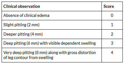
The degree of edema is commonly described on a 0 to 4+ scale in order of increasing severity.[42] One clinical assessment method uses the following scoring system:[45]
[Figure caption and citation for the preceding image starts]: A clinical assessment scoring systemCreated by BMJ Knowledge Centre based on Brodovicz KG, McNaughton K, Uemura N, et al. Reliability and feasibility of methods to quantitatively assess peripheral edema. Clin Med Res. 2009 Jun;7(1-2):21-31. [Citation ends].
Describing the level that edema extends to proximally on the limbs is also useful in communicating the severity of peripheral edema. However, these descriptions are inexact and may fluctuate depending on the patient's positioning.
Ankle circumference is a simple quantitative test for clinical practice with good inter-rater reliability. It is measured 7 cm proximal to the medial malleolus.[45]
Stemmer sign is present if the examiner cannot pinch a fold of skin at the base of the patient's second toe. It is a sensitive test for primary and secondary lymphedema.[46] Lymphedema is pitting in the early stages and becomes nonpitting due to adipose deposition and fibrosis when the lymphedema is chronic and advanced.
A key discriminating factor in narrowing etiologies is whether the edema is limited to a single extremity or is generalized. Generalized edema suggests a systemic illness such as heart failure, cirrhosis, or nephrotic syndrome. Localized edema suggests a localized venous or lymphatic abnormality.
Comprehensive physical examination is crucial, with particular focus on the cardiac and pulmonary systems.
Examination findings that might indicate an underlying cause for peripheral edema include:
Cardiovascular: additional S3 or S4 heart sounds, gallop rhythm, raised jugular venous pressure in heart failure; pulsus paradoxus, pericardial friction rub, muffled heart sounds, and elevated jugular venous pressure in pericardial disease; Kussmaul sign (rise in jugular venous pressure on inspiration), S3 heart sound, mitral regurgitation murmur, elevated jugular venous pressure in restrictive cardiomyopathy; systolic murmur at the left lower sternal border and prominent ventricular impulse in the left parasternal region in tricuspid regurgitation.
Respiratory: rales if there is pulmonary edema, wheeze in cor pulmonale caused by COPD.
Abdominal: splenomegaly, ascites, jaundice, spider nevi, and enlarged periumbilical veins (caput medusae) in cirrhosis; tender hepatomegaly in Budd-Chiari syndrome, hepatomegaly and ascites in heart failure, restrictive cardiomyopathy or tricuspid regurgitation; palpable mass in lower abdomen may indicate tumor causing external pressure on pelvic veins.
Dermatologic: presence of varicose veins, fibrosis, ulcers, or skin changes (such as hemosiderin staining, atrophie blanche, and venous stasis dermatitis) suggests a chronic venous insufficiency. Lymphatic insufficiency often coexists with venous insufficiency. Calf asymmetry, warmth, tenderness, and erythema, dilated superficial veins and/or palpable cords behind the leg, occur in DVT. Periorbital edema is characteristic of nephrotic syndrome. Dry skin and coarse hair occur in myxedema.
Musculoskeletal: severe pain on passive stretch in compartment syndrome, disproportionate to the injury severity. In compartment syndrome, pulse exam may be normal, and unless an artery is injured, the capillaries refill. Impaired calf muscle pump function; may be associated with ankle foot orthoses or drop foot.
General: tachypnea, tachycardia, altered mental status, hypotension, decreased urine output, diarrhea, warm flushed or cold clammy skin in sepsis. Obesity, large tonsils, short neck with large circumference suggest obstructive sleep apnea.
[Figure caption and citation for the preceding image starts]: Atrophie blanche and hemosiderin deposition in a patient with chronic venous insufficiencyFrom the collection of Dr Joseph L. Mills; used with permission [Citation ends].
Diagnostic testing
The underlying etiology of edema can be suggested by basic screening tests.[6][47]
Serum chemistry, hepatic function panel, serum protein, albumin, and urinalysis are indicated. Thyroid-stimulating hormone levels, cardiac troponins, B-type natriuretic peptide (BNP)/N-terminal pro-B-type natriuretic peptide (NT-proBNP), ECG, chest x-ray and/or echocardiography may be useful when some features of history or physical exam suggest endocrine or cardiopulmonary disease.
Evaluation of lymphatic drainage with lymphoscintigraphy may be considered when clinical assessment and investigations have excluded systemic and local vascular factors as a cause of peripheral edema. Lymphoscintigraphy is rarely used in the routine investigation of peripheral edema.
[Figure caption and citation for the preceding image starts]: Approach to the patient with peripheral edema. DVT, deep vein thrombosis; HTN, hypertensionCreated by Ethan Cumbler, MD [Citation ends].
Asymmetric edema
For patients with asymmetric peripheral edema and no apparent cause (such as radiation or surgical lymph node dissection), testing should focus on proximal venous or lymphatic obstruction.
A duplex ultrasound of the inferior vena cava or iliac vessels should be considered if a proximal cause is suspected, and can identify venous insufficiency, venous thrombosis, or nonthrombotic iliac vein lesions (e.g., May-Thurner syndrome [compression of the left common iliac vein by the overlying right common iliac artery]).
Ultrasound may identify hypoechoic regions superficial to the mid calf muscles if a popliteal fossa cyst has ruptured. Pelvic ultrasound may reveal presence of a mass causing preferential compression of venous flow from one lower extremity.
Computed tomography or magnetic resonance imaging may be considered if pelvic or abdominal disease is suspected.
If history arouses suspicion of filariasis then blood smears for detection of microfilariae are indicated.
Genetic testing may be ordered in patients with primary lymphedema, although familial/syndromic causes are rare.[48][49][50]
Bilateral edema
For patients with diffuse or bilateral edema, the initial diagnostic focus is on determining whether an organ system is in dysfunction.
Serum protein and albumin can screen for the etiologies that cause hypoproteinemia, such as nephrotic syndrome, protein-losing enteropathies, or profound malnutrition. Low protein is also a feature of cirrhosis. If low serum albumin is detected, urinalysis and serum creatinine are ordered to determine if the cause is renal protein wasting, while liver function tests and coagulation studies are ordered to assess hepatic synthetic dysfunction.
Normal urine and hepatic testing suggests malnutrition or protein-losing enteropathy as the cause of hypoproteinemia. The history should distinguish between these two clinical entities.
Serum chemistry, blood urea nitrogen, and creatinine should be ordered if there is suspicion of renal failure, heart failure, or cirrhosis. Elevated creatinine and reduced glomerular filtration rate indicate renal disease, while hyponatremia is a common finding in patients with heart failure or cirrhosis.
Thyroid-stimulating hormone levels may be indicated as part of an initial screen, especially if other features of the history and physical examination suggest hypothyroidism.
ECG and a chest x-ray are advisable to evaluate for evidence of cardiac congestion, especially when the physical examination demonstrates elevated jugular venous pressure or abnormal cardiopulmonary examination findings.
If abnormalities are found on ECG and chest x-ray, then echocardiography is indicated. Echocardiography can establish right or left heart dysfunction as a cause of congestive features or can suggest pulmonary hypertension as a result of pulmonary disease or sleep-disordered breathing.
Measurement of BNP or NT-proBNP is also useful in the diagnosis or exclusion of heart failure.[51][52] Follow-up testing evaluates the underlying reason for the cardiac or pulmonary dysfunction.
Lymphoscintigraphy
Lymphoscintigraphy can objectively distinguish lymphatic pathology from other causes of peripheral edema; however, it is rarely used in the routine investigation of peripheral edema.
Lymphoscintigraphy involves peripheral intradermal or subcutaneous injection of a radiolabeled tracer. Images are recorded with a gamma camera to demonstrate the flow of the tracer through the lymphatic system.
Signs of lymphatic dysfunction include delayed, asymmetric, or absent violation of regional lymph nodes, and "dermal backflow" (presence of radiolabeled tracer in the dermal lymphatic channels).[53] A normal result shows symmetric transport of the tracer through discrete lymph nodes.[54] Lymphoscintigraphy has a 96% sensitivity and 100% specificity for lymphedema.[55]
How to take a venous blood sample from the antecubital fossa using a vacuum needle.
How to record an ECG. Demonstrates placement of chest and limb electrodes.
Use of this content is subject to our disclaimer

