History and exam
Key diagnostic factors
common
Gottron papules
Pathognomonic of DM.
Violaceous to dusky-red flat-topped papules and plaques over the dorsal surface of knuckles (and more rarely the wrists, elbows, knees, and malleoli).[107] The surface may be slightly scaling or sometimes psoriasiform.
Telangiectasia may develop within the lesions.[Figure caption and citation for the preceding image starts]: Macular erythematous patches (Gottron papules) over the dorsal surface of the hands, especially over the metacarpophalangeal and proximal interphalangeal jointsAdapted from BMJ Case Reports 2009 [doi:10.1136/bcr.06.2009.2027] Copyright © 2009 by the BMJ Publishing Group Ltd [Citation ends].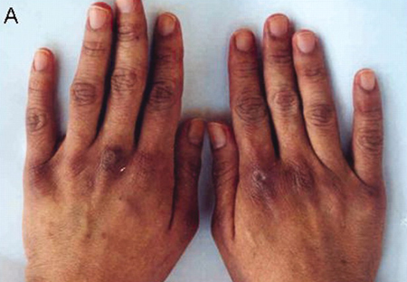 [Figure caption and citation for the preceding image starts]: Macular erythematous patches (Gottron papules) over the elbowsAdapted from BMJ Case Reports 2009 [doi:10.1136/bcr.06.2009.2027] Copyright © 2009 by the BMJ Publishing Group Ltd [Citation ends].
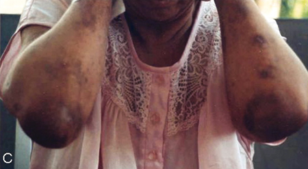 [Figure caption and citation for the preceding image starts]: Macular erythematous patches (Gottron papules) over the kneesAdapted from BMJ Case Reports 2009 [doi:10.1136/bcr.06.2009.2027] Copyright © 2009 by the BMJ Publishing Group Ltd [Citation ends].
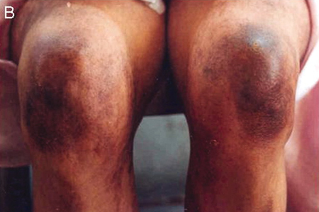
heliotrope rash with or without periorbital edema
macular violaceous erythema
Symmetric violaceous erythematous macular rash with characteristic distributions:[108][109][110]
Gottron sign: macular patches over knuckles, elbows, and knees.
V-sign: distribution in "v" of neck and upper chest indicating photodistribution.
Shawl sign: distribution in nape of neck, posterior shoulders, and upper back.
Holster sign: distribution on lateral thigh or hip.
Linear extensor erythemas: involving extensor surfaces of legs, thighs, upper arms, forearms, dorsal fingers, and feet.
Facial rash can also be seen in malar distribution with perioral sparing.
Erythema involving the scalp is often associated with pruritus.[Figure caption and citation for the preceding image starts]: Macular erythematous patches (Gottron papules) over the dorsal surface of the hands, especially over the metacarpophalangeal and proximal interphalangeal jointsAdapted from BMJ Case Reports 2009 [doi:10.1136/bcr.06.2009.2027] Copyright © 2009 by the BMJ Publishing Group Ltd [Citation ends].
periungual erythema, nail-fold capillary dilation, cuticular overgrowth
Commonly seen, although also present in other connective tissue disorders.[70][71]
Dilated capillary loops with vascular dropout and cuticular overgrowth (ragged cuticles) can be indicators of disease activity.[Figure caption and citation for the preceding image starts]: Macular erythematous patches (Gottron papules) over the toes with dystrophic cuticlesAdapted from BMJ Case Reports 2009 [doi:10.1136/bcr.06.2009.2027] Copyright © 2009 by the BMJ Publishing Group Ltd [Citation ends].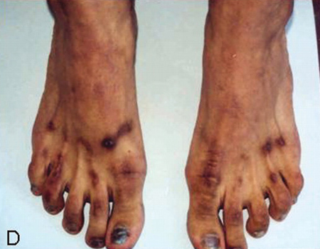
"mechanic's" hands
Hyperkeratosis, scaling and fissuring of palms of hands and palmar aspect of fingers.[45]
May be part of the antisynthetase syndrome.
proximal muscle weakness
The onset of muscle weakness is usually subacute. Weakness is proximal and symmetric (e.g., difficulty getting out of a chair, climbing stairs, washing/combing hair, and lifting arms).
Distal motor involvement is present in only 10% to 20% of cases and is more likely in later disease.[73]
Other diagnostic factors
common
photosensitivity
poikiloderma vasculare atrophicans
Nonspecific sign.
Atrophic areas with varying hypopigmentation and hyperpigmentation, telangiectasia, and scaling.
May occur in areas of chronic macular violaceous erythema.
pruritus
May be a prominent symptom, especially with scalp involvement. Its presence may help differentiate between DM and cutaneous lupus.[108]
fatigue and malaise
Common but nonspecific symptom.
dyspnea
weight loss
May be reported along with other constitutional symptoms. Significant weight loss, however, is not common and when present should raise the possibility of malignancy-associated DM.
uncommon
fever
Nonspecific symptom.
myalgia
Present in approximately 30% of patients.[3]
arthralgia
dysphagia
Present in 15% to 50% of patients, esophageal disease is a poor prognostic sign.
Proximal dysphagia due to involvement of pharyngeal and esophageal striated muscle correlates with severity of muscle involvement elsewhere and responds to treatment.
Distal dysphagia is due to nonstriated muscle disease and is more common in patients with overlap syndromes, especially in overlap with systemic sclerosis.[110]
palpitations and syncope
Cardiac involvement occurs in 50% of patients but is rarely symptomatic unless disease is advanced.[79] Arrhythmias are the most common cardiac abnormality and these may be symptomatic.[80] Cardiac failure due to cardiac myositis is rare.
Clinically apparent cardiac involvement is a poor prognostic indicator.[77][78]
cutaneous calcinosis
Rare in adult patients but present in approximately 30% to 70% of children with DM.[48][68][69]
Subcutaneous, firm, yellow- or flesh-colored nodules, often over bony prominences.
Complications include extrusion with secondary infection.
Calcinosis in juvenile DM can be extensive, resulting in contractures and loss of function.
erythroderma
Total-body erythema is a rare manifestation.
vesiculobullous lesions
May be more common in malignancy-associated DM.
leukocytoclastic vasculitis
Rare manifestation. When present suggests malignancy-associated DM.[113]
cutaneous necrosis
Rare manifestation. When present suggests malignancy-associated DM.[114]
nonscarring alopecia
When present may be a sign of disease flare.[110]
Risk factors
strong
genetic predisposition
Suggested by the occurrence in monozygotic twins, familial cases, and familial association with other autoimmune diseases.[14][15][16]
There are reported associations between DM/polymyositis (PM) and HLA-DRB1*03 in white people and HLA-DRB1*14 in Koreans.[19][20] HLA-DRB1*09:01 and HLA-DRB1*12:01 alleles may contribute to susceptibility to adult DM in the Han Chinese population.[17]
HLA-DQA1 0501 is reported to be associated with juvenile DM and tumor necrosis factor 308A polymorphism with photosensitivity in DM.[18][21]
bimodal age distribution: children and age >40 years
ultraviolet radiation
The rash is often photodistributed, developing in sun-exposed areas. Some patients report sensitivity to sunlight, although others do not make this association.[58]
Increased ultraviolet-light-induced apoptotic cells in the skin may lead to suprathreshold concentration of antigenic peptides.[59] Ultraviolet-radiation intensity has been shown to be a strong contributor to DM and is related to the proportion of anti-Mi-2 antibodies.[22][23][24]
weak
infectious agents
Direct muscle invasion by bacteria and viruses causing myositis is well recognized. Although no good evidence is available for infectious agents as environmental triggers, some findings may support this hypothesis.[13]
There are some reports of DM/polymyositis developing in patients with antibodies to Borrelia burgdorferi and Toxoplasma gondii.[25][26]
High titers of coxsackievirus antibodies have been found in cases of idiopathic inflammatory myopathies early in disease course.[27][28] Picornavirus-like particles have been found in muscle biopsies of patients with myositis.[29]
drugs
Drugs that have been implicated in causing DM include D-penicillamine, hydroxyurea, statins, phenylbutazone, and terbinafine.[36][37][38][39][40][41][42][43][44]
The most commonly reported are statins (HMG-CoA reductase inhibitors), including simvastatin, lovastatin, and fluvastatin.[39][40][41][42] These reports are rare and statins are more likely to be associated with myalgia and minor elevations of creatine kinase (CK) rather than myositis per se.
Use of this content is subject to our disclaimer