The key to diagnosis of an ACL injury is a thorough history and physical exam.
History
Key historical features indicating an ACL injury include a high-risk mechanism (contact or noncontact), an audible pop at the time of injury, rapid development of hemarthrosis, inability to return to play, pain, and a feeling that the knee is unstable or buckles with attempted weight bearing.[42]American Academy of Orthopaedic Surgeons. Management of anterior cruciate ligament injuries: evidence-based clinical practice guideline. Aug 2022 [internet publication].
https://www.aaos.org/globalassets/quality-and-practice-resources/anterior-cruciate-ligament-injuries/aclcpg.pdf
[45]Shelbourne KD, Rowdon GA. Anterior cruciate ligament injury: the competitive athlete. Sports Med. 1994;17:132-40.
http://www.ncbi.nlm.nih.gov/pubmed/8171223?tool=bestpractice.com
History of prior ACL injury and female sex should also heighten suspicion.[11]Hootman JM, Dick R, Agel J. Epidemiology of collegiate injuries for 15 sports: summary and recommendations for injury prevention initiatives. J Athl Train. 2007 Apr-Jun;42(2):311-9.
https://www.ncbi.nlm.nih.gov/pmc/articles/PMC1941297
http://www.ncbi.nlm.nih.gov/pubmed/17710181?tool=bestpractice.com
[12]Mihata LC, Beutler AI, Boden BP. Comparing the incidence of anterior cruciate ligament injury in collegiate lacrosse, soccer, and basketball players. Am J Sports Med. 2006;34:899-904.
http://www.ncbi.nlm.nih.gov/pubmed/16567461?tool=bestpractice.com
[13]Agel J, Arendt EA, Bershadsky B. Anterior cruciate ligament injury in National Collegiate Athletic Association basketball and soccer: a 13 year review. Am J Sports Med. 2005;33:524-30.
http://www.ncbi.nlm.nih.gov/pubmed/15722283?tool=bestpractice.com
[14]Myer GD, Ford KR, Hewett TE. Methodological approaches and rationale for training to prevent anterior cruciate ligament injuries in female athletes. Scand J Med Sci Sports. 2004;14:275-85.
http://www.ncbi.nlm.nih.gov/pubmed/15387801?tool=bestpractice.com
[15]Giugliano DN, Solomon JL. ACL tears in female athletes. Phys Med Rehabil Clin N Am. 2007;18:417-38.
http://www.ncbi.nlm.nih.gov/pubmed/17678760?tool=bestpractice.com
[28]Faude O, Junge A, Kindermann W, et al. Risk factors for injuries in elite female soccer players. Br J Sports Med. 2006;40:785-90.
http://www.ncbi.nlm.nih.gov/pmc/articles/PMC2564395
http://www.ncbi.nlm.nih.gov/pubmed/16825269?tool=bestpractice.com
Patients are often adolescents and young adults, or middle-aged individuals who are involved in extensive exercise.
Physical examination
Physical exam usually reveals a hemarthrosis if the injury was recent. Chronic ACL tears may or may not have an associated effusion. Acutely, range of motion is usually impaired, especially in flexion. This may be due to a combination of pain and stiffness from hemarthrosis, as well as associated bone bruise, meniscal tears, or articular cartilage injury.[46]Maffulli N, Binfield PM, King JB, et al. Acute haemarthrosis of the knee in athletes: a prospective study of 106 cases. J Bone Joint Surg Br. 1993;75:945-9.
http://www.bjj.boneandjoint.org.uk/content/jbjsbr/75-B/6/945.full.pdf
http://www.ncbi.nlm.nih.gov/pubmed/8245089?tool=bestpractice.com
Tenderness is found at the lateral femoral condyle and lateral tibial plateau.[Figure caption and citation for the preceding image starts]: T1-weighted MRI showing ACL tearFrom the personal collection of Philip H. Cohen [Citation ends].
It is crucial to perform tests gently and smoothly, and to have the patient relax; otherwise the patient's discomfort and fear may lead to guarding, tensing of the hamstrings, and false-negative or inconclusive results.
Lachman test
The Lachman test is the most accurate maneuver for detecting an acute ACL tear, whereas the pivot shift test performs better in chronic tears or under anesthesia.[42]American Academy of Orthopaedic Surgeons. Management of anterior cruciate ligament injuries: evidence-based clinical practice guideline. Aug 2022 [internet publication].
https://www.aaos.org/globalassets/quality-and-practice-resources/anterior-cruciate-ligament-injuries/aclcpg.pdf
[47]Ostrowski JA. Accuracy of 3 diagnostic tests for anterior cruciate ligament tears. J Athl Train. 2006;41:120-1.
http://www.ncbi.nlm.nih.gov/pmc/articles/PMC1421494
http://www.ncbi.nlm.nih.gov/pubmed/16619105?tool=bestpractice.com
[48]Benjaminse A, Gokeler A, van der Schans CP. Clinical diagnosis of an anterior cruciate ligament rupture: a meta-analysis. J Orthop Sports Phys Ther. 2006;36:267-88.
http://www.ncbi.nlm.nih.gov/pubmed/16715828?tool=bestpractice.com
The Lachman test is performed with the patient in a supine position. The patient’s knee is positioned at 20 to 30 degrees of flexion. One hand is placed on the patient’s thigh and the other behind the tibia, with the clinician's thumb on the tibial tuberosity. The tibia is then pulled anteriorly. If the ACL is intact, a firm end point is felt. If this does not occur, and there is >2 mm of anterior movement compared with the uninjured knee, the test is positive, suggesting a torn ACL. [Figure caption and citation for the preceding image starts]: Lachman maneuverFrom the personal collection of Philip H. Cohen [Citation ends].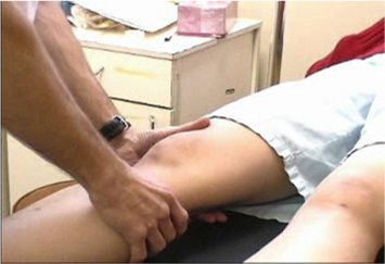
Anterior drawer test
The anterior drawer test will often be positive, but is less sensitive and specific.[49]Solomon DH, Simel DL, Bates DW, et al. The rational clinical examination. Does this patient have a torn meniscus or ligament of the knee? Value of the physical examination. JAMA. 2001;286:1610-20.
http://www.ncbi.nlm.nih.gov/pubmed/11585485?tool=bestpractice.com
This test involves placing the patient in a supine position and flexing the hips to 45 degrees, with the knees at 90 degrees and the patient's feet on the table.[Figure caption and citation for the preceding image starts]: Anterior and posterior drawer test starting positionFrom the personal collection of Philip H. Cohen [Citation ends].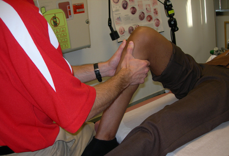 Sitting on the patient's feet, the clinician takes hold of the tibia and pulls it anteriorly. If the tibia moves more than usual, the test is positive.
Varus/valgus stress test
A standard part of the physical exam.[42]American Academy of Orthopaedic Surgeons. Management of anterior cruciate ligament injuries: evidence-based clinical practice guideline. Aug 2022 [internet publication].
https://www.aaos.org/globalassets/quality-and-practice-resources/anterior-cruciate-ligament-injuries/aclcpg.pdf
Although not specific for identifying ACL tears, this test is necessary to evaluate for other or concomitant injuries.[Figure caption and citation for the preceding image starts]: Varus and valgus stability testingFrom the personal collection of Philip H. Cohen [Citation ends].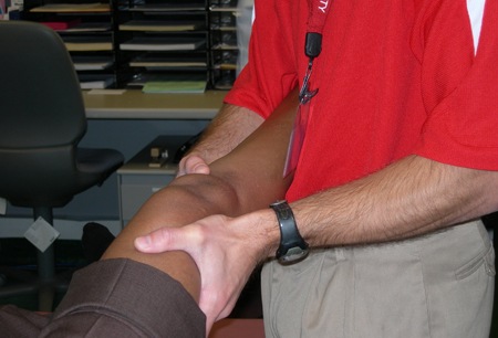
Pivot shift maneuver
Usually performed under anesthesia in the operating room, before or after reconstruction surgery. It is technically difficult, but the best test for dynamic rotatory instability.[50]Kocher MS, Steadman JR, Briggs KK, et al. Relationships between objective assessment of ligament stability and subjective assessment of symptoms and function after anterior cruciate ligament reconstruction. Am J Sports Med. 2004;32:629-34.
http://www.ncbi.nlm.nih.gov/pubmed/15090377?tool=bestpractice.com
Arthrocentesis
Usually not indicated, but sometimes done for symptom relief if tense hemarthrosis is present, or to assist with the diagnosis in confusing cases.
Imaging
Ottawa knee rules recommend x-rays in acute knee injury in an adult if any of the following features is present:[51]Stiell IG, Wells GA, McDowell I, et al. Use of radiography in acute knee injuries: need for clinical decision rules. Acad Emerg Med. 1995 Nov;2(11):966-73.
https://onlinelibrary.wiley.com/doi/epdf/10.1111/j.1553-2712.1995.tb03123.x
http://www.ncbi.nlm.nih.gov/pubmed/8536122?tool=bestpractice.com
[52]Expert Panel on Musculoskeletal Imaging, Taljanovic MS, Chang EY, et al. ACR appropriateness criteria® acute trauma to the knee. J Am Coll Radiol. 2020 May;17(5s):S12-S25.
https://www.doi.org/10.1016/j.jacr.2020.01.041
http://www.ncbi.nlm.nih.gov/pubmed/32370956?tool=bestpractice.com
Inability to bear weight at time of injury and at time of evaluation
Isolated tenderness at patella or fibular head
Active flexion of knee <90 degrees
Age >55 years.
X-rays are usually negative, but may reveal the lateral capsular sign/Segond fracture; a small capsular avulsion off the lateral aspect of the proximal tibia.[53]Davis DS, Post WR. Segond fracture: lateral capsular ligament avulsion. J Orthop Sports Phys Ther. 1997;25:103-6.
http://www.ncbi.nlm.nih.gov/pubmed/9007767?tool=bestpractice.com
This is uncommon but virtually pathognomonic for ACL tear.
Magnetic resonance imaging (MRI)
MRI has excellent sensitivity and specificity for ACL tears, and can reveal associated injuries. However, MRI is not necessary if the clinical diagnosis is clear.[54]Thomas S, Pullagura M, Robinson E, et al. The value of magnetic resonance imaging in our current management of ACL and meniscal injuries. Knee Surg Sports Traumatol Arthrosc. 2007;15:533-6.
http://www.ncbi.nlm.nih.gov/pubmed/17225179?tool=bestpractice.com
[Figure caption and citation for the preceding image starts]: T2-weighted MRI showing bone bruise, effusion, and flattening of sulcus terminalisFrom the personal collection of Philip H. Cohen [Citation ends].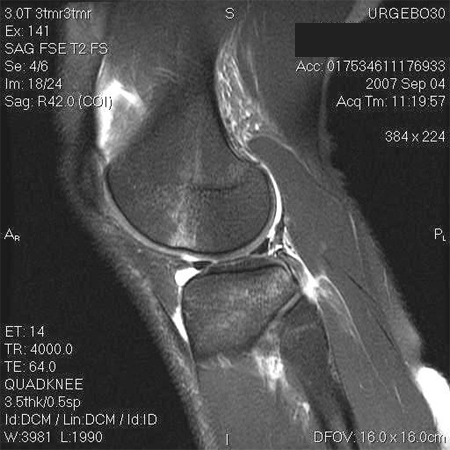
MRI may be useful diagnostically when the clinical exam is impaired by factors such as patient guarding, tense effusion, or a locked joint (intra-articular block from displaced meniscal tear, etc.). MRI can also be helpful when evaluating the possibility of a torn ACL graft.[55]Bining J, Andrews G, Forster BB. The ABCs of the anterior cruciate ligament: a primer for magnetic resonance imaging assessment of the normal, injured and surgically repaired anterior cruciate ligament. Br J Sports Med. 2009;43:856-62.
http://www.ncbi.nlm.nih.gov/pubmed/19864590?tool=bestpractice.com
MRI is often ordered as an initial test, especially in high-level athletes. However, this practice is generally unnecessary and is not supported by evidence-based research. In particular, if the patient is a child, do not order MRI until all appropriate clinical, laboratory, and plain radiographic exams have been completed. MRI is costly, frequently requires sedation in the young child and, if needed, is best ordered by the specialist who will be treating the patient.[56]American Academy of Pediatrics – Section on Orthopaedics and the Pediatric Orthopaedic Society of North America. Five things physicians and patients should question. Choosing Wisely, an initiative of the ABIM Foundation. 2022 [internet publication].
https://web.archive.org/web/20230209025014/https://www.choosingwisely.org/societies/american-academy-of-pediatrics-section-on-orthopaedics-and-the-pediatric-orthopaedic-society-of-north-america
From a surgical perspective, there is some evidence that preoperative MRI may be helpful in determining the suitability of a given patient's patellar tendon for use in an ACL bone-patellar tendon-bone autograft.[57]Chang CB, Seong SC, Kim TK. Preoperative magnetic resonance assessment of patellar tendon dimensions for graft selection in anterior cruciate ligament reconstruction. Am J Sports Med. 2009;37:376-82.
http://www.ncbi.nlm.nih.gov/pubmed/19036719?tool=bestpractice.com
CT is usually not appropriate unless there has been significant trauma to the knee, or a fall or acute trauma to the knee and no fracture or tibial plateau fracture is seen on radiographs, or suspected occult fracture or suspected additional bone or soft tissue injury.[52]Expert Panel on Musculoskeletal Imaging, Taljanovic MS, Chang EY, et al. ACR appropriateness criteria® acute trauma to the knee. J Am Coll Radiol. 2020 May;17(5s):S12-S25.
https://www.doi.org/10.1016/j.jacr.2020.01.041
http://www.ncbi.nlm.nih.gov/pubmed/32370956?tool=bestpractice.com
However, if the patient is a child, do not order CT until all appropriate clinical, laboratory, and plain radiographic exams have been completed in order to limit radiation exposure.[56]American Academy of Pediatrics – Section on Orthopaedics and the Pediatric Orthopaedic Society of North America. Five things physicians and patients should question. Choosing Wisely, an initiative of the ABIM Foundation. 2022 [internet publication].
https://web.archive.org/web/20230209025014/https://www.choosingwisely.org/societies/american-academy-of-pediatrics-section-on-orthopaedics-and-the-pediatric-orthopaedic-society-of-north-america