Treatment algorithm
Please note that formulations/routes and doses may differ between drug names and brands, drug formularies, or locations. Treatment recommendations are specific to patient groups: see disclaimer
reduction and immobilization ± surgical referral
Once the diagnosis has been confirmed, reduction should be attempted. For a successful outcome, adequate analgesia and sedation are necessary before the reduction procedure is attempted. There are numerous reduction maneuvers for shoulder injuries, which are usually performed under local anesthesia (i.e., intra-articular lidocaine) combined with procedural sedation (e.g., intravenous morphine, midazolam, or etomidate).
[ ![]() ]
]
The choice for sedation depends on the treating physician and must be accompanied by continuous monitoring of the patient with capnography and pulse oximetry, as well as frequent blood pressure measurements.
Local anesthesia on its own should be reserved for patients with contraindications to procedural sedation.
Each of the reduction methods works by abduction and external rotation to disengage the humeral head from the glenoid, with axial traction to reduce it. Irrespective of the technique used, the physician should feel a distinct clunk as the shoulder reduces.
The arm should be immobilized and placed in a sling or a sling and swathe.
An anteroposterior (AP) and lateral radiograph should be obtained to confirm reduction of the humeral head, and to ensure that no iatrogenic fractures have occurred during the reduction.[74][Figure caption and citation for the preceding image starts]: Normal axillary x-ray view of a reduced shoulder dislocation, showing congruency of the glenohumeral jointPersonal collection of Dr Paul Novakovich [Citation ends].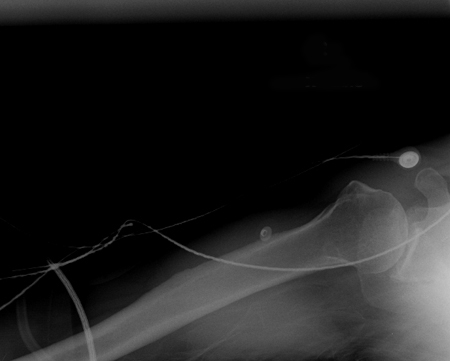
Once the patient is alert, it is important to perform a neurologic and vascular exam.
The patient should wear the sling for approximately 3 weeks.
Patients under 25 years of age should be referred to an orthopedic surgeon for consideration of further intervention (i.e., possible arthroscopic or open repair), as this age group is at significant risk for recurrence. Long-term data support primary stabilization via anatomic Bankart repair (over simple arthroscopic lavage or nonoperative treatment) for young, high-risk patients with a first-time shoulder dislocation.[75][76] One systematic review found that patients, particularly active men in their 20s and 30s, undergoing treatment for a first-time anterior shoulder dislocation with a surgical stabilization procedure, can be expected to experience significantly lower rates of recurrent instability and a significantly decreased need for a future stabilization procedure when compared with patients treated nonoperatively.[77]
The Latarjet procedure is a commonly used approach for managing chronic and recurrent anterior shoulder dislocation, especially in the presence of bone loss.[78] It involves transplant of the coracoid process to the scapular neck and has demonstrated excellent long-term clinical outcomes and return to sport rate.[79] It may be more effective than Bankart repair for recurrent instability of the shoulder.[80] Both open and arthroscopic Latarjet procedures result in significantly improved function and outcome in patients with anterior shoulder instability.[81] However, the Latarjet procedure has been associated with a complication rate of 15% to 30%; specific complications include graft-related issues (11.7%), hardware-related complications (6.5%), nerve injuries (0.7% to 4%), recurrent instability (8%), and revision (5%).[78]
reduction and immobilization
Reduction is usually performed using local anesthesia (i.e., intra-articular lidocaine) combined with procedural sedation (e.g., intravenous morphine, midazolam, or etomidate). The choice for sedation depends on the treating physician and must be accompanied by continuous monitoring of the patient with capnography and pulse oximetry, as well as frequent blood pressure measurements.
Local anesthesia on its own should be reserved for patients with contraindications to procedural sedation.
The patient should be supine on the bed with the physician positioned on the affected side with an assistant close to the head of the bed. In young children, management of radial head subluxation/dislocation (nursemaid elbow) may be more effective and less painful when performed with the arm in pronation as opposed to supination.[87]
[ ![]() ]
]
The arm should be initially extended to 30° flexion.
The overall gross alignment of the elbow is then manipulated so that the olecranon appears centered between the medial and lateral condyle of the humerus.
The forearm is then slowly flexed to approximately 90° with the physician providing longitudinal traction to the forearm while the assistant provides countertraction to the patient's humerus.
The arm is then flexed even further with direct downward pressure applied to the olecranon.[44]
If reduction is successful, the physician should feel an audible clunk as the elbow is reduced.
It is important not to flex the arm forcefully if there is significant resistance because the coronoid process is typically perched on the distal humerus. Forceful flexion without adequate traction can cause a fracture of this structure, which will result in future instability.
Upon reduction, the arm is placed in a posterior splint at 90° flexion with neutral rotation of the forearm.[44]
An anteroposterior (AP) and lateral plain film radiograph of the elbow should be obtained to ensure that the joint is concentrically reduced. [Figure caption and citation for the preceding image starts]: Anteroposterior x-ray view of a reduced elbow dislocationPersonal collection of Dr Paul Novakovich [Citation ends].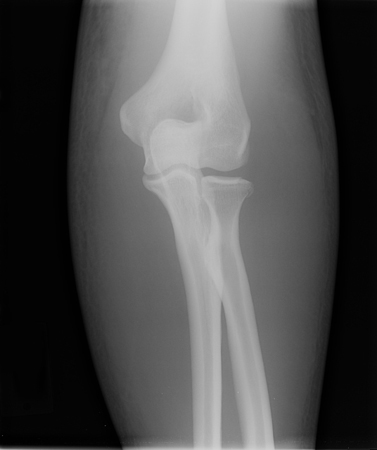
Once the patient is alert, it is important to perform a neurologic and vascular exam.
Choose a patient group to see our recommendations
Please note that formulations/routes and doses may differ between drug names and brands, drug formularies, or locations. Treatment recommendations are specific to patient groups. See disclaimer
Use of this content is subject to our disclaimer