Patients with significant brachial plexus injuries (those that persist beyond a few hours or days) are at risk for permanent loss of function if the injury is not managed rapidly and efficiently.
The two main factors that direct management are the speed of nerve regeneration (about 1 mm per day; 1 inch per month) and the time beyond which motor recovery is impossible (about 1 year from date of injury).[18]Shin AY, Spinner RJ, Steinmann SP, et al. Adult traumatic brachial plexus injuries. J Am Acad Orthop Surg. 2005;13:382-96.
http://www.ncbi.nlm.nih.gov/pubmed/16224111?tool=bestpractice.com
[29]Nath RK, Mackinnon SE. Nerve transfers in the upper extremity. Hand Clin. 2000;16:131-9.
http://www.ncbi.nlm.nih.gov/pubmed/10696582?tool=bestpractice.com
These parameters also determine the distance from the nerve root beyond which motor recovery is unlikely in severe injuries: 30 cm (12 inches). Because the brachial plexus is usually injured at very proximal levels, the effect of these inflexible rules is to render lower root injuries (physically farther from their motor targets than the upper roots) devastating in most cases.[11]Kim DH, Cho YJ, Tiel RL, et al. Outcomes of surgery in 1019 brachial plexus lesions treated at Louisiana State University Health Sciences Center. J Neurosurg. 2003;98:1005-16.
http://www.ncbi.nlm.nih.gov/pubmed/12744360?tool=bestpractice.com
Upper root injuries may be severe but are usually reconstructable if treated by appropriate specialists.[1]Nath RK, Lyons AB, Bietz G. Physiological and clinical advantages of median nerve fascicle transfer to the musculocutaneous nerve following brachial plexus root avulsion injury. J Neurosurg. 2006;105:1-5.
http://www.ncbi.nlm.nih.gov/pubmed/17405252?tool=bestpractice.com
[30]Leechavengvongs S, Witoonchart K, Uerpairojkit C, et al. Combined nerve transfers for C5 and C6 brachial plexus avulsion injury. J Hand Surg (Am). 2006;31:183-9.
http://www.ncbi.nlm.nih.gov/pubmed/16473676?tool=bestpractice.com
Physical and occupational therapy, including appropriate splints and braces, are useful prior to any surgical intervention and even if surgery is not done. The general principles of therapy include prevention and treatment of stiffness and contracture formation (especially in the hand), electrical stimulation, and sensory re-education where applicable.
Pain management
Management of pain should involve a multidisciplinary team that includes pain specialists. Neuropathic pain is commonly associated with brachial plexus injury.[31]Lovaglio AC, Socolovsky M, Di Masi G, et al. Treatment of neuropathic pain after peripheral nerve and brachial plexus traumatic injury. Neurol India. 2019 Jan-Feb;67(Suppl):S32-7.
https://www.neurologyindia.com/article.asp?issn=0028-3886;year=2019;volume=67;issue=7;spage=32;epage=37;aulast=Lovaglio
http://www.ncbi.nlm.nih.gov/pubmed/30688230?tool=bestpractice.com
When usual analgesia is not effective, specific treatments for neuropathic pain, such as gabapentin, carbamazepine, tricyclic antidepressants, topical lidocaine or capsaicin, or opioids (e.g., oxycodone), may be used. The anti-inflammatory drug treatment celecoxib has been shown to improve sciatic functional index (SFI) significantly in rats, following sciatic nerve crush injury. Celecoxib may be considered in the treatment of concomitant peripheral nerve injuries, if present.[32]Cámara-Lemarroy CR, Guzmán-de la Garza FJ, Barrera-Oranday EA, et al. Celecoxib accelerates functional recovery after sciatic nerve crush in the rat. J Brachial Plex Peripher Nerve Inj. 2008;3:25.
http://www.ncbi.nlm.nih.gov/pmc/articles/PMC2607269
http://www.ncbi.nlm.nih.gov/pubmed/19036161?tool=bestpractice.com
Upper (C5-6) and middle (C7) root injuries
The consequences of injury at this level depend on severity of the inciting event, whether mechanical, neoplastic, or inflammatory. Loss of function of the shoulder and biceps due to a complete C5-6 injury renders the limb severely disabled, but capable of some useful function since the hand is intact. The physiologic parameters that result in permanent paralysis by 1 year after injury dictate management. An electromyogram can give useful information about injury patterns and the presence of avulsion but is not very specific as to lesser degrees of injury: a neuropraxic injury may mimic a complete rupture.[17]Smith, SJM. Electrodiagnosis. In: Birch R, Bonney G, Wynn Parry CB, eds. Surgical disorders of the peripheral nerves. London: Churchill-Livingstone; 1998:467-90. The end result is that serial clinical examination tends to be the best method to indicate surgery versus conservative management. Most surgeons will evaluate the patient for reconstructive surgery if there is no evidence of motor recovery by 4 to 6 months after injury.[1]Nath RK, Lyons AB, Bietz G. Physiological and clinical advantages of median nerve fascicle transfer to the musculocutaneous nerve following brachial plexus root avulsion injury. J Neurosurg. 2006;105:1-5.
http://www.ncbi.nlm.nih.gov/pubmed/17405252?tool=bestpractice.com
[30]Leechavengvongs S, Witoonchart K, Uerpairojkit C, et al. Combined nerve transfers for C5 and C6 brachial plexus avulsion injury. J Hand Surg (Am). 2006;31:183-9.
http://www.ncbi.nlm.nih.gov/pubmed/16473676?tool=bestpractice.com
[33]Martin E, Senders JT, DiRisio AC, et al. Timing of surgery in traumatic brachial plexus injury: a systematic review. J Neurosurg. 2018 May 1;1-13.
https://www.doi.org/10.3171/2018.1.JNS172068
http://www.ncbi.nlm.nih.gov/pubmed/29999446?tool=bestpractice.com
[34]Noland SS, Bishop AT, Spinner RJ, et al. Adult traumatic brachial plexus injuries. J Am Acad Orthop Surg. 2019 Oct 1;27(19):705-16.
https://journals.lww.com/jaaos/Fulltext/2019/10010/Adult_Traumatic_Brachial_Plexus_Injuries.1.aspx
http://www.ncbi.nlm.nih.gov/pubmed/30707114?tool=bestpractice.com
This allows time for nerve regeneration down to the paralyzed muscles within the critical 1-year time frame.
Nerve transfer techniques are the treatment of choice for microsurgical repair.[1]Nath RK, Lyons AB, Bietz G. Physiological and clinical advantages of median nerve fascicle transfer to the musculocutaneous nerve following brachial plexus root avulsion injury. J Neurosurg. 2006;105:1-5.
http://www.ncbi.nlm.nih.gov/pubmed/17405252?tool=bestpractice.com
[21]Garg R, Merrell GA, Hillstrom HJ, et al. Comparison of nerve transfers and nerve grafting for traumatic upper plexus palsy: a systematic review and analysis. J Bone Joint Surg Am. 2011;93:819-29.
http://www.ncbi.nlm.nih.gov/pubmed/21543672?tool=bestpractice.com
[22]Wells ME, Gonzalez GA, Childs BR, et al. Radial to axillary nerve transfer outcomes in shoulder abduction: a systematic review. Plast Reconstr Surg Glob Open. 2020 Sep 23;8(9):e3096.
https://www.ncbi.nlm.nih.gov/pmc/articles/PMC7544396
http://www.ncbi.nlm.nih.gov/pubmed/33133948?tool=bestpractice.com
[23]Schessler MJ, McClellan WT. The role of nerve transfers for C5-C6 brachial plexus injury in adults. W V Med J. 2010 Jan-Feb;106(1):12-7.
http://www.ncbi.nlm.nih.gov/pubmed/20088304?tool=bestpractice.com
[24]Lanier ST, Hill JR, James AS, et al. Approach to the pan-brachial plexus injury: variation in surgical strategies among surgeons. Plast Reconstr Surg Glob Open. 2020 Nov 24;8(11):e3267.
https://www.ncbi.nlm.nih.gov/pmc/articles/PMC7722554
http://www.ncbi.nlm.nih.gov/pubmed/33299725?tool=bestpractice.com
Factors that influence extent of clinical improvement include patient age, mechanism of injury, timing of surgery, and multiple nerve transfers versus single nerve transfers.[22]Wells ME, Gonzalez GA, Childs BR, et al. Radial to axillary nerve transfer outcomes in shoulder abduction: a systematic review. Plast Reconstr Surg Glob Open. 2020 Sep 23;8(9):e3096.
https://www.ncbi.nlm.nih.gov/pmc/articles/PMC7544396
http://www.ncbi.nlm.nih.gov/pubmed/33133948?tool=bestpractice.com
Nerve transfer requires specialized knowledge and experience, so nerve grafting techniques may be used by nonspecialist surgeons.[35]Gkiatas I, Papadopoulos D, Korompilias A, Vekris M, Beris A, Kostas-Agnantis I. Traumatic upper plexus palsy: is the exploration of brachial plexus necessary? Eur J Orthop Surg Traumatol. 2019 Feb;29(2):255-62.
http://www.ncbi.nlm.nih.gov/pubmed/30483967?tool=bestpractice.com
If the patient has passed the 1-year deadline for primary reconstruction, terminal muscle atrophy is present and provision of a nerve supply cannot result in function. However, microsurgical techniques can provide both nerve (nerve transfer) and viable new muscle (free muscle transfer) to the limb such that at least functional elbow flexion is achievable.[24]Lanier ST, Hill JR, James AS, et al. Approach to the pan-brachial plexus injury: variation in surgical strategies among surgeons. Plast Reconstr Surg Glob Open. 2020 Nov 24;8(11):e3267.
https://www.ncbi.nlm.nih.gov/pmc/articles/PMC7722554
http://www.ncbi.nlm.nih.gov/pubmed/33299725?tool=bestpractice.com
[34]Noland SS, Bishop AT, Spinner RJ, et al. Adult traumatic brachial plexus injuries. J Am Acad Orthop Surg. 2019 Oct 1;27(19):705-16.
https://journals.lww.com/jaaos/Fulltext/2019/10010/Adult_Traumatic_Brachial_Plexus_Injuries.1.aspx
http://www.ncbi.nlm.nih.gov/pubmed/30707114?tool=bestpractice.com
[36]Hoang D, Chen VW, Seruya M. Recovery of elbow flexion after nerve reconstruction versus free functional muscle transfer for late, traumatic brachial plexus palsy: a systematic review. Plast Reconstr Surg. 2018 Apr;141(4):949-59.
http://www.ncbi.nlm.nih.gov/pubmed/29595730?tool=bestpractice.com
Partial ulnar nerve transfer (PUNT) and intercostal nerve transfer (ICNT) have been shown to be equally effective for reconstructing elbow flexion in patients with upper brachial plexus injuries.[37]Kakinoki R, Ikeguchi R, Dunkan SF, et al. Comparison between partial ulnar and intercostal nerve transfers for reconstructing elbow flexion in patients with upper brachial plexus injuries. J Brachial Plex Peripher Nerve Inj. 2010;5:4.
http://www.ncbi.nlm.nih.gov/pmc/articles/PMC2881072
http://www.ncbi.nlm.nih.gov/pubmed/20181014?tool=bestpractice.com
Shoulder function is achievable with early nerve reconstructive techniques, but after 6 to 8 months shoulder fusion is ultimately a better solution than nerve repair.[38]Chammas M, Goubier JN, Coulet B, et al. Glenohumeral arthrodesis in upper and total brachial plexus palsy. A comparison of functional results. J Bone Joint Surg (Br). 2004;86:692-5.
http://www.ncbi.nlm.nih.gov/pubmed/15274265?tool=bestpractice.com
[Figure caption and citation for the preceding image starts]: Repair of avulsed spinal nerveFrom the collection of the Texas Nerve and Paralysis Institute, Dr Rahul Nath, Founder and Medical Director; used with permission [Citation ends].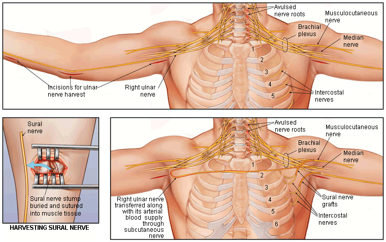
Lower root (C8-T1) injuries and total (C5-T1) injuries
Due to the time and distance constraints of nerve regeneration discussed above, severe injuries to the lower roots usually result in difficult reconstructive situations. The best solution tends to be a staged combination of nerve transfer to restore innervation, followed several months later by free muscle transfer to replace atrophic muscle for hand function. In complete injuries to all 5 roots, there are few ipsilateral sources for nerve restoration, but intercostal nerves can serve as partial donors.[39]Merrell GA, Barrie KA, Katz DL, et al. Results of nerve transfer techniques for restoration of shoulder and elbow function in the context of a meta-analysis of the English literature. J Hand Surg (Am). 2001;26:303-14.
http://www.ncbi.nlm.nih.gov/pubmed/11279578?tool=bestpractice.com
The contralateral C7 nerve is then available to provide a source of motor as well as sensory nerve for reconstruction of injured limb function.
Once the intercostal and contralateral C7 nerves have been transferred, multiple free muscle transfers are needed to restore elbow flexion and finger function.[40]Doi K, Muramatsu K, Hattori Y, et al. Restoration of prehension with the double free muscle technique following complete avulsion of the brachial plexus. Indications and long-term results. J Bone Joint Surg Am. 2000;82:652-66.
http://www.ncbi.nlm.nih.gov/pubmed/10819276?tool=bestpractice.com
Shoulder movement is restored by glenohumeral fusion (although fusion seems intuitively to prevent movement, in actuality this fusion links the paralyzed humerus to the still-active scapula, thus allowing arm movement through shrugging and other scapular movements that will still be present).[38]Chammas M, Goubier JN, Coulet B, et al. Glenohumeral arthrodesis in upper and total brachial plexus palsy. A comparison of functional results. J Bone Joint Surg (Br). 2004;86:692-5.
http://www.ncbi.nlm.nih.gov/pubmed/15274265?tool=bestpractice.com