Approach
The presentation of groin pain in relation to physical activity can lead to a number of very different diagnoses. Careful attention to the history and examination findings can guide the clinician in determining the correct diagnosis and/or further workup if indicated.
History
Onset of pain (acute versus chronic), severity, and radiating features, if present, should be described.
A history of trauma, mechanism of injury, aggravating and/or alleviating factors, occupational status, recreational activities, and constitutional symptoms, if present, should all be ascertained.
Physical examination
The precise location of pain (groin, pubis, iliac crest, greater trochanter, thigh, buttock, sacroiliac joint, abdomen, lumbar spine) should be indicated by the patient without the examiner's assistance. Careful inspection and palpation of pertinent anatomic structures with reproduction of pain on palpation should be noted.
Careful clinical examination, including a number of functional tests of the relevant muscles, tendons, and joints, should be done, including:[14][15]
Squeeze test for the adductors
Palpation and modified Thomas test for the iliopsoas
Examination of the inguinal canal and conjoint tendon for incipient hernia
Impingement and apprehension test for the hip joints.
Gait pattern should be observed, in particular:
Trendelenburg gait, which results from insufficient strength in the gluteus medius[Figure caption and citation for the preceding image starts]: Positive Trendelenburg signFrom the collection of Cedric J. Ortiguera, MD [Citation ends].
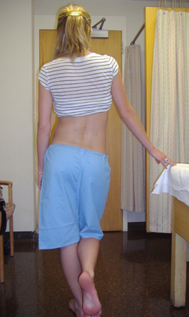
Coxalgic gait, which results from the patient quickly unloading the painful leg while bearing weight.
Passive and active range of motion of the ipsilateral hip and knee should be performed to identify painful or limited range of motion, and compared with the contralateral side.
Examination of the abdomen, lower back, or genitourinary system should be performed at the examiner's discretion when pain referred to the hip from other areas is suspected.
Stress fracture
Patients may have a history leading to suspicion of overuse injury (e.g., endurance athlete or military recruit).
Standard x-rays may not reveal fractures. Patients should have an MRI if there is a high clinical suspicion, even if no fracture is visible on plain x-rays.
Soft-tissue injury
The Doha consensus classification of groin injuries in athletes are in principle followed in this topic.[1]
Adductor-related groin pain
History of athletic overuse or traumatic injury.
Examination characterized by pain with palpation and pain against resisted adduction of the hip.
Usually a clinical diagnosis, although ultrasound or MRI may be helpful in showing injury to the tendon or the enthesis.
Iliopsoas-related groin pain
History of athletic overuse or traumatic injury.
Examination characterized by pain with palpation of the lower part and the tendon of the iliopsoas.
Usually a clinical diagnosis; ultrasound or MRI can be helpful showing widening of the tendon.
Inguinal-related groin pain (also known as incipient hernia, sports hernia, or athletic pubalgia)
Caused by a weakening of the posterior inguinal wall.
Pain typically reproducible with palpation of the conjoint tendon at the pubic tubercle, at the external opening of the inguinal ring, or performing oblique sit-ups.
Ultrasound is often useful to demonstrate the soft posterior wall; MRI might be used to rule out other conditions with similar presentations.
Bursitis
Typically chronic overuse injury in recreational or competitive athlete.
Point tenderness with palpation on greater trochanter seen with trochanteric bursitis.
Iliopsoas bursitis is not very common and cannot be diagnosed clinically because of the surrounding soft-tissue envelope; may demonstrate pain with active hip flexion, worse with resistance.
Usually not a clinical diagnosis; ultrasound or MRI are the definitive tests.
Osteitis pubis
Speculative diagnosis describing the changes seen in the pubic bone and around the pubic symphysis with x-ray and MRI. Commonly seen in soccer players. These changes probably reflect the amount of stress inflicted by sport. It is not a definite sign of pathology, but rather a sign of a repair reaction.
Typically concomitant with adductor-related pain, and presents with activity-related pain in the lower abdomen and groin region (unilateral or bilateral).
May be difficult to distinguish from adductor injury, and the 2 entities can exist concomitantly.
Plain film radiographs may show widening of the symphysis, irregular articular surfaces, and/or sclerosis.
MRI shows marrow edema. One study has shown bone marrow edema, as demonstrated by MRI, around the pubic symphysis in the pubic bone to be nearly as common in soccer players with no symptoms of groin pain (31%) as in soccer players suffering from long-standing groin pain (51%).[8] Bone marrow edema is not diagnostic of groin injury.
Labral tears
Degenerative labral tears seen in 93% of cadaveric specimens.[16]
Associated with twisting or pivoting activities, such as ballet, football, or soccer.
May present with mechanical symptoms of catching or clicking.
Pain typically reproducible on examination with anterior impingement test (hip flexion, adduction, and internal rotation – FADIR tests).
Plain x-rays demonstrate the underlying bony abnormality (e.g., dysplasia, femoral acetabular impingement) in the majority of cases.
MRI arthrogram is the most sensitive noninvasive test available to identify labral tears.[17]
Diagnostic intra-articular injections with an anesthetic and/or corticosteroid may be of value in trying to determine the etiology of pain as being intra-articular (labral tear, chondral lesion) or extra-articular (tendinitis/bursitis, muscular, intra-abdominal, lumbar).
Use of this content is subject to our disclaimer