Images and videos
Images
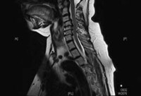

Degenerative cervical spine disease
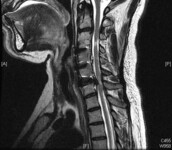
Cervical MRI (sagittal T2) with moderate degenerative joint disease but no significant spinal cord compression
Dennis A. Turner, MA, MD
See this image in context in the following section/s:

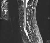
Degenerative cervical spine disease
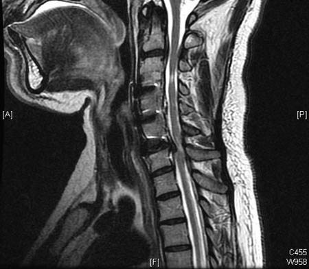
Cervical MRI (sagittal T2) with mild degenerative joint disease and disk bulging
Dennis A. Turner, MA, MD
See this image in context in the following section/s:

Degenerative cervical spine disease
Chart showing average dermatome size and location. Radicular pain is usually confined to a single dermatome
From Gray's Anatomy of the Human Body (29th ed., US); used with permission
See this image in context in the following section/s:

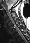
Degenerative cervical spine disease
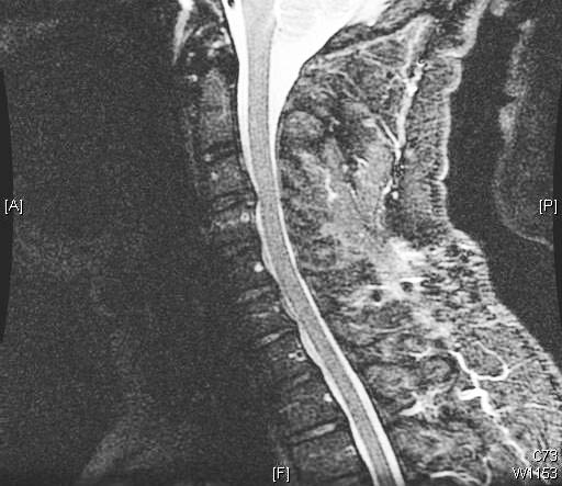
A single level of spinal cord compression with T2 changes, on cervical sagittal T2 sequence in the presence of symptomatic degenerative cervical myelopathy
Dennis A. Turner, MA, MD
See this image in context in the following section/s:

Degenerative cervical spine disease
Diagram of subsets of cervical spondylosis, including various symptoms possibly arising within the larger field of asymptomatic (radiographic) spondylosis
Dennis A. Turner, MA, MD
See this image in context in the following section/s:
Degenerative cervical spine disease
Previous spinal cord compression at C3/4 on sagittal T2 MRI, with residual T2 changes, and new compression at C2/3 and C6/7, with T2 changes
Dennis A. Turner, MA, MD
See this image in context in the following section/s:

Degenerative cervical spine disease
Severe, multilevel degenerative disk disease changes but without significant spinal cord compression (i.e., neither deformation nor intrinsic T2 changes) on cervical MRI (sagittal T2)
Dennis A. Turner, MA, MD
See this image in context in the following section/s:
Use of this content is subject to our disclaimer