Approach
The approach to the patient with hyponatremia involves using a combination of clinical assessment and measurements of serum osmolality and urinary sodium. The cause is often apparent from the history and examination, but other conditions can only be diagnosed with the use of targeted investigations. The most common causes of euvolemic hyponatremia are medications and syndrome of inappropriate antidiuretic hormone secretion (SIADH).[24][25] SIADH is a diagnosis of exclusion, characterized by a euvolemic clinical picture with a low urine output and increased urine osmolality. By contrast, cerebral salt-wasting syndrome produces a hypovolemic clinical picture, with a high urine output and normal or low urine osmolality. However, there are no clearly defined thresholds, and controversy exists as to whether the distinction between these two conditions is possible or meaningful.
A rapid decline in serum sodium levels over 24 to 48 hours can lead to severe cerebral edema and central nervous system symptoms including headache, muscle cramps, reversible ataxia, psychosis, lethargy, apathy, anorexia, and agitation. This is an acute medical emergency. If no acute intervention is initiated to increase sodium level, patients can develop coma, brainstem herniation, and respiratory arrest, leading to death.
A slower decline in sodium levels over several days or weeks is usually asymptomatic; when it is symptomatic, it produces milder cerebral edema, which does not lead to brainstem herniation.
Establishing the type of hyponatremia
The first step is to measure effective serum osmolality (tonicity). [ Osmolality Estimator (serum) Opens in new window ] If it is normal (275-295 mOsm/kg H₂O), the patient has pseudohyponatremia (isotonic hyponatremia), which is an artifact produced by high serum lipid or protein levels. Newer electrodes measure sodium directly and the measured sodium concentration will be normal if these electrodes are used. The most common cause of high protein levels is multiple myeloma; this diagnosis is already known in the majority of patients.
If the serum osmolality is >295 mOsm/kg H₂O, the patient has redistributive hyponatremia (hypertonic), which is either due to hyperglycemia or to the absorption or administration of a hypertonic fluid (e.g., mannitol, glycine, or sorbitol).
Hyperglycemia is usually caused by diabetes but can also be caused by medications (beta-blockers, thiazide diuretics, corticosteroids, niacin, pentamidine, protease inhibitors, some antipsychotics) or by stress from a recent stroke, myocardial infarction, trauma, infection, or inflammation. A fasting or random serum glucose measurement establishes hyperglycemia as the cause. The serum HbA1c is elevated in people with poorly controlled diabetes and may also be useful. Medication-induced hyponatremia and hyperglycemia should resolve once the causative agent is discontinued.
Hypertonic hyponatremia due to mannitol, glycine, or sorbitol is usually easily established by examination of fluids administered intravenously or fluids used to irrigate the operative field during transurethral resection of prostate or hysteroscopy. However, it can be confirmed by calculation of the serum osmolar gap. [ Osmolal gap calculator Opens in new window ] A difference >10 indicates the presence of nonsodium effective osmoles such as mannitol, glycine, or sorbitol.
If the serum osmolality is <275 mOsm/kg H₂O, the patient has hypotonic hyponatremia (hypovolemic, euvolemic, or hypervolemic).
Patients with hypovolemic hyponatremia will have signs of volume depletion (decreased skin turgor, reduced jugular venous pressure, decreased blood pressure).
Patients with hypervolemic hyponatremia will have an elevated jugular venous pressure and peripheral edema.
The absence of any of these signs indicates that the patient is euvolemic.
Because hyponatremia can arise in hypervolemic, euvolemic, and hypovolemic states, hyponatremia and its cause may not initially be clear.[26] The most important test to identify the etiology in patients with hypovolemic hypotonic hyponatremia, euvolemic hypotonic hyponatremia, or hypervolemic hypotonic hyponatremia is measurement of urinary sodium. A spot urinary sodium test is available that allows urinary sodium to be quickly and conveniently measured in a random urine sample.
Hypovolemic hyponatremia: urinary sodium >20 mEq/L indicates renal sodium losses and urinary sodium ≤20 mEq/L indicates extrarenal sodium losses.
Hypervolemic hyponatremia: urinary sodium >20 mEq/L suggests acute kidney injury or chronic kidney disease and a urinary sodium ≤20 mEq/L suggests edematous disorders such as heart failure, cirrhosis, or nephrotic syndrome.
Patients with euvolemic hyponatremia always have a urinary sodium >20 mEq/L.[Figure caption and citation for the preceding image starts]: Algorithm for the diagnosis of hypotonic hyponatremia. SIADH, syndrome of inappropriate antidiuretic hormoneProduced by the BMJ Knowledge Centre [Citation ends].
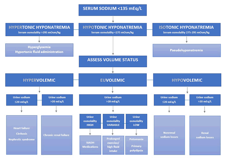
It is also important to measure urine osmolality. Urine osmolality is <100 mOsm/kg H₂O in cases of excessive water intake, but >100 mOsm/kg H₂O in all other causes. However, in general, urine osmolality is measured primarily to assess disease severity and is not useful for elucidating the underlying cause. It should be noted that causes such as endocrinopathies (glucocorticoid deficiency), potassium depletion, and diuretic use may present with either a euvolemic or hypovolemic state depending on the severity of the disease.
Hypovolemic hyponatremia with urinary sodium >20 mEq/L
Renal disease
Salt-wasting nephropathy should be considered as a cause in all patients with hypovolemic hyponatremia with a urinary sodium >20 mEq/L. Many patients will have a known diagnosis or a positive family history of tubulointerstitial disease (interstitial nephritis, medullary cystic kidney disease, partial urinary tract obstruction, and polycystic kidney disease). Salt-wasting nephropathy often precedes the onset of renal failure in these conditions. An abdominal mass is often present in polycystic kidney disease. Patients with medullary cystic kidney disease have early signs of severe anemia such as pallor.
The serum creatinine may be normal or elevated with a normal or reduced glomerular filtration rate. Urinalysis reveals hematuria and/or proteinuria, depending on the underlying cause. Renal ultrasound will detect obstruction, hydronephrosis, kidney stones, or cysts. Contrast-enhanced abdominal CT scanning is a more definitive imaging tool for assessing the number, size, and location of the cysts in polycystic kidney disease and medullary cystic kidney disease. Genetic testing is the definitive method for distinguishing these conditions. Renal biopsy is required for the definitive diagnosis of interstitial nephritis, and should be considered in consultation with a renal specialist.
Central nervous system cause
If there is a history of recent head injury, intracranial surgery, subarachnoid hemorrhage, stroke, or brain tumors, cerebral salt-wasting syndrome should be considered as the cause. These conditions can also cause SIADH, but SIADH causes euvolemic hyponatremia and is a diagnosis of exclusion. A complete history and central nervous system examination should identify the cause.
A CT scan brain will identify signs of hemorrhage or skull fractures. An MRI brain is the preferred modality to detect intracranial tumors and to assess ischemic stroke once hemorrhagic stroke has been excluded by CT.
Mineralocorticoid deficiency
Should also be excluded as a cause. Symptoms and signs are usually nonspecific and include nausea, vomiting, myalgia, arthralgia, and clinical signs of volume depletion.
Serum potassium is usually elevated. A decreased morning serum cortisol is diagnostic. A decreased cortisol response to adrenocorticotropic hormone is seen.
Hypovolemic hyponatremia with urinary sodium ≤20 mEq/L
This is produced by inappropriate replacement of extrarenal sodium and fluid losses with hypotonic fluids. This may be caused by replacement of excessive sweating (often due to prolonged exercise in a hot environment) by oral tap water or by intravenous hypotonic fluids. These causes are evident from the history and examination of fluid charts.
Other causes of fluid loss that may prompt inappropriate fluid replacement include vomiting, diarrhea, gastrointestinal fistulas or drainage tubes, and third spacing of fluids caused by peritonitis, pancreatitis, burns, or small bowel obstruction.
Hypervolemic hyponatremia with urinary sodium ≤20 mEq/L
Congestive heart failure
A history of myocardial infarction should prompt consideration of congestive heart failure. Symptoms include fatigue, decreased exercise tolerance, dyspnea on exertion, orthopnea, and paroxysmal nocturnal dyspnea. Clinical signs include edema, displaced cardiac apex, hepatojugular reflux, jugular venous distension, S3 gallop, pulmonary rales, and hepatomegaly.
Chest x-ray may show cardiomegaly, pulmonary edema, or a pleural effusion.
Measurement of natriuretic peptide biomarker (B-type [BNP] and N-terminal pro B-type [NT-proBNP]) levels is helpful to rule in or exclude heart failure in patients with a suspected cardiac cause of dyspnea.[27]
An ECG may show anterior Q waves (indicating a previous myocardial infarction), bundle branch block, atrial arrhythmias, ventricular arrhythmias, left axis deviation, or left ventricular hypertrophy. An echocardiogram detects systolic and diastolic dysfunction. Valve lesions, signs of pericardial injury, or cardiomyopathy may also be seen.
Liver cirrhosis
A history of alcohol misuse, intravenous drug use, unprotected intercourse, obesity, blood transfusion, or known hepatitis infection should prompt suspicion of cirrhosis. Cirrhosis severe enough to cause hypervolemic hyponatremia is usually symptomatic. Symptoms include fatigue, weakness, weight gain, and pruritus. Signs include edema, jaundice, ascites, collateral circulation, hepatosplenomegaly, leukonychia, palmar erythema, spider angiomata, telangiectasia, jaundiced sclera, hepatic fetor, and altered mental status.
LFTs are abnormal, and the pattern depends on the cause of cirrhosis.
An abdominal ultrasound can be used to detect signs of advanced cirrhosis such as liver surface nodularity, small liver, possible hypertrophy of left/caudate lobe, ascites, splenomegaly, and increased diameter of the portal vein (≥13 mm) or collateral vessels. Liver biopsy provides a definitive diagnosis, but is only necessary if the diagnosis cannot be established based on clinical features, investigations, and imaging.
Nephrotic syndrome
Should be suspected if there is a history of long-standing diabetes, malignancy, systemic lupus erythematosus, HIV infection, multiple myeloma, connective tissue diseases, or amyloidosis, or use of known causative medications (pamidronate, lithium, gold, penicillamine, or nonsteroidal anti-inflammatory drugs, and, very rarely, interferon alfa, heroin, mercury, or formaldehyde). Patients present with leg or generalized edema and foamy urine. Patients may also have Muehrcke lines (due to hypoalbuminemia) or xanthelasmas (due to hypertriglyceridemia).
Serum albumin levels are low. The plasma creatinine may be normal or elevated depending on the stage of disease. A 24-hour urine collection for protein shows nephrotic range proteinuria (>3 g/24 hours). Renal biopsy is required for the definitive diagnosis of many of the underlying causes, and should be considered in consultation with a renal specialist.
Hypervolemic hyponatremia with urinary sodium >20 mEq/L
Indicates renal disease with impaired sodium excretion. In most patients the diagnosis is already known, but further assessment is required if a new diagnosis of renal disease is made. Signs and symptoms of renal disease may be present and include jaundice, skin bruising, poor concentration/memory, or myoclonus.
Serum creatinine is elevated with a reduced glomerular filtration rate, and urinalysis reveals hematuria and/or proteinuria depending on the underlying cause. Renal ultrasound can be useful to assess the cause, and may reveal small kidneys, obstruction or hydronephrosis, and kidney stones. A kidney biopsy is required for the definitive diagnosis of intrinsic causes of renal disease, and should be considered in consultation with a renal specialist.
Euvolemic hyponatremia
Thiazide diuretics
The hyponatremia may appear within days or even years of starting the medication, and will resolve once thiazide diuretics have been discontinued.[14]
Excessive oral fluid intake
If a history of schizophrenia or psychotic depression is present, psychogenic polydipsia should be considered. A history of polydipsia and polyuria is present. Patients usually complain of a persistent sensation of dry mouth. This may sometimes be due to phenothiazine medications and sometimes be due to the underlying condition. The clinical examination is usually normal, although weight gain due to high water intake may occur in extreme cases. The water intake is so excessive that it overwhelms the capacity of the kidney to resorb water, and urine osmolality is <100 mOsm/kg H₂O.
If a history of chronic alcohol misuse is present, potomania should be considered. The CAGE questionnaire can be helpful in identifying patients with alcohol misuse. CAGE alcohol questionnaire Opens in new window A CAGE score >2 is suspicious. Potomania is precipitated by drinking more than 6 liters of beer a day on a background of chronic poor dietary intake. Urine osmolality is <100 mOsm/kg H₂O. Serum bilirubin levels are typically elevated, and aspartate aminotransferase and alanine aminotransferase are rarely >200 units/L.
Postoperative period
A transient increase in antidiuretic hormone secretion occurs during this period. The hyponatremia is self-limiting. However, administration of hypotonic fluids during this time can produce a more severe or prolonged hyponatremia.
Large volumes of hypertonic fluids (glycine, mannitol, or sorbitol) are used to irrigate the operative field during transurethral resection of the prostate or hysteroscopy. If the fluid is absorbed without the solute, this can produce euvolemic hypotonic hyponatremia.
Large volumes of hypotonic fluids are used to irrigate the operative field during endometrial ablation. If absorbed, this can produce severe acute hyponatremia.
SIADH
If all other causes of euvolemic hyponatremia have been excluded, the patient has SIADH.[28]
Known drug causes include vasopressin, nonsteroidal anti-inflammatory drugs, nicotine, chlorpropamide, carbamazepine, tricyclic antidepressants, selective serotonin reuptake inhibitors, vincristine, thioridazine, cyclophosphamide, clofibrate, and ecstasy (MDMA) use.
Recent head injury, intracranial surgery, subarachnoid hemorrhage, stroke, brain tumors, meningitis, or brain abscess can cause SIADH.
A history of cough, shortness of breath, or pleuritic chest pain should prompt consideration of respiratory causes of SIADH. These include pneumonia, lung abscess, COPD, cystic fibrosis, and positive pressure ventilation.
Ectopic ADH secretion by tumors is an important cause to exclude. The most common source is small cell lung cancer; other cancers are rare causes. These include cervical cancer, lymphoma, leukemia, and pancreatic adenoma.
Appropriate investigations depend on the cause identified by the clinical features. If there is no identifiable cause, the patient is diagnosed to have idiopathic SIADH.

How to take a venous blood sample from the antecubital fossa using a vacuum needle.
How to insert a peripheral intravascular catheter into the dorsum of the hand.
Use of this content is subject to our disclaimer