Images and videos
Images


Acute pyelonephritis
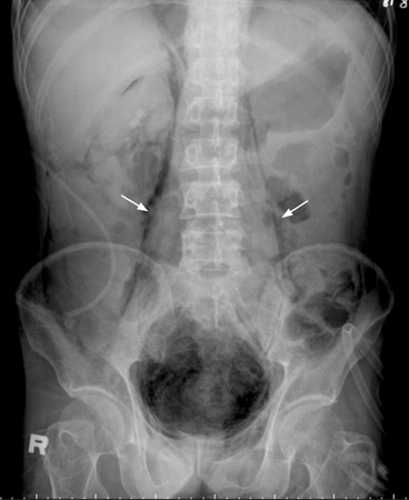
Psoas muscle shadow enhanced by retroperitoneal air in emphysematous pyelonephritis
Courtesy of Shih-Hung Tsai and Shi-Jye Chu, Department of Emergency Medicine, Tri-Service General Hospital, National Defense Medical Center, Taiwan, Republic of China
See this image in context in the following section/s:
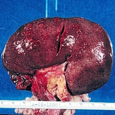
Acute pyelonephritis
Cross-section of a kidney with acute suppurative pyelonephritis. The white streaks are purulent exudates throughout the kidney, in both the tubules and the interstitium
McLay RN, Harrison JH, Fermin CD, et al. Tulane gross pathology tutorial. Tulane University School of Medicine; 1997
See this image in context in the following section/s:
Acute pyelonephritis
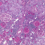
Interstitial infiltrates and edema in acute pyelonephritis. Some glomeruli also show evidence of a second kidney disease, focal glomerulosclerosis
Courtesy of Dr Jean L. Olson, Department of Pathology, University of California, San Francisco, CA
See this image in context in the following section/s:

Acute pyelonephritis
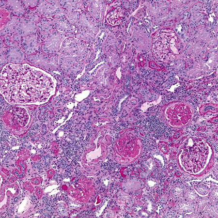
Higher magnification: polymorphonuclear cell infiltrates in and around the renal tubules
Courtesy of Dr Jean L. Olson, Department of Pathology, University of California, San Francisco, CA
See this image in context in the following section/s:
Acute pyelonephritis
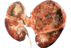
External surface of the kidney with multifocal irregular, whitish, raised lesions consisting of purulent exudates
McLay RN, Harrison JH, Fermin CD, et al. Tulane gross pathology tutorial. Tulane University School of Medicine; 1997
See this image in context in the following section/s:
Use of this content is subject to our disclaimer