Images and videos
Images


Evaluation of acute abdomen
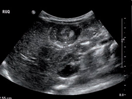
Cholecystitis: Ultrasound of acute cholecystitis and presence of gallstones
From the collection of Dr Charles Bellows
See this image in context in the following section/s:
Evaluation of acute abdomen
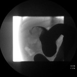
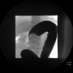
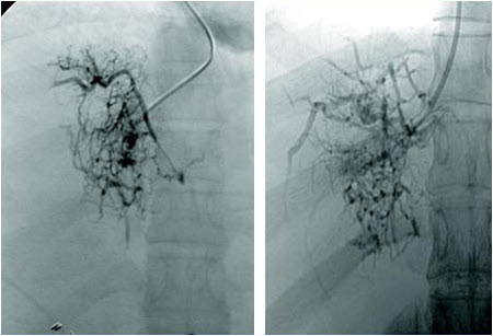
Budd-Chiari syndrome: Hepatic venogram demonstrating "spider web" and thrombus in the inferior vena cava
Liver Transplantation Journal. 2006 Nov;12(11 suppl 2):S21-2; reprinted with permission of John Wiley & Sons, Inc
See this image in context in the following section/s:

Evaluation of acute abdomen
Intussusception: Site of intussusception as revealed by abdominal x-ray, showing the meniscus
From the collection of Dr David J. Hackam
See this image in context in the following section/s:

Evaluation of acute abdomen
Liver Abscess: Gross pathology of amebic abscess of liver; tube of "chocolate" pus from abscess
CDC/Dr Mae Melvin; Dr. E. West of Mobile, AL
See this image in context in the following section/s:

Evaluation of acute abdomen
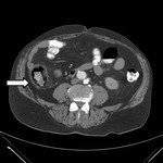
Crohn disease: CT scan demonstrating thickening of the terminal ileum in a patient with Crohn disease exacerbation
Provided by Drs Wissam Bleibel, Bishal Mainali, Chandrashekhar Thukral, and Mark A. Peppercorn
See this image in context in the following section/s:

Evaluation of acute abdomen
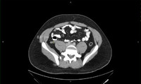
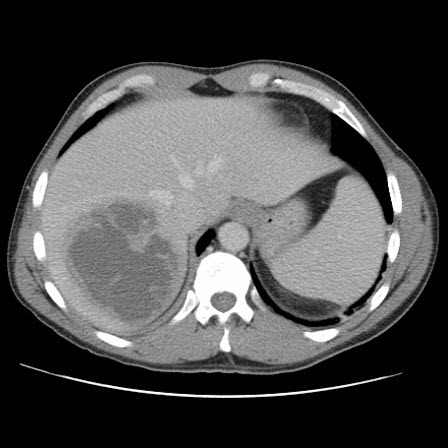
Abdominal aortic aneurysm: CT scan of a ruptured abdominal aortic aneurysm
University of Michigan, specifically the cases of Dr Upchurch reflecting the Departments of Vascular Surgery and Radiology
See this image in context in the following section/s:

Evaluation of acute abdomen
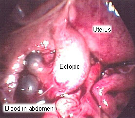
Ectopic pregnancy: Surgical extraction of ectopic pregnancy
From the personal collection of Dr Melissa Fries, Washington Hospital Center; used with permission
See this image in context in the following section/s:
Evaluation of acute abdomen
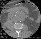
Liver abscess: CT scan (coronal view) showing liver abscess in a 46-year-old man who presented with fever, fatigue, and cough
From the collection of Massachusetts General Hospital radiology images
See this image in context in the following section/s:

Evaluation of acute abdomen
Liver abscess: CT scan showing a liver abscess (7 cm x 5 cm) in a 46-year-old man who presented with fever, fatigue, and cough
From the collection of Massachusetts General Hospital radiology images
See this image in context in the following section/s:

Evaluation of acute abdomen
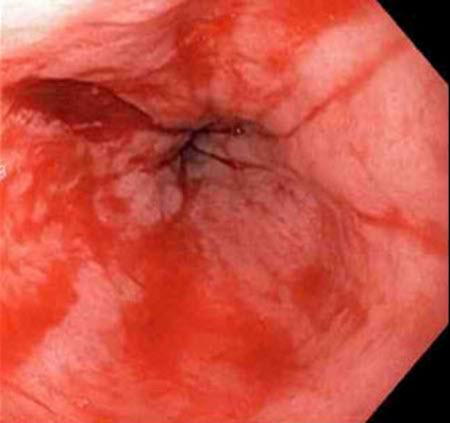
Mallory-Weiss tear: Nonbleeding adherent clot
From the collection of Juan Carlos Munoz, MD, University of Florida
See this image in context in the following section/s:

Evaluation of acute abdomen
Endometriosis: Laparoscopic image of ovarian endometrioma
From the collection of Dr Jonathon Solnik; used with permission
See this image in context in the following section/s:

Evaluation of acute abdomen
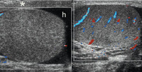
Testicular torsion: Bilateral transverse color Doppler images in a 12-year-old boy with right-sided scrotal pain of sudden onset, showing no color flow signals in the right testis, which is enlarged and has heterogeneous echogenicity; reactive hydrocele (h) and thickening of the scrotal wall (*) are also seen; testicular torsion and bell clapper deformity were confirmed at surgery
Aso C, et al. Radiographics. 2005 Sep-Oct;25(5):1197-214; used with permission
See this image in context in the following section/s:
Evaluation of acute abdomen
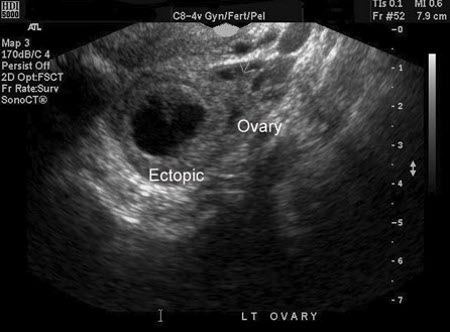
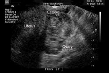
Ectopic pregnancy: Ultrasound image of ectopic pregnancy
From the personal collection of Dr Melissa Fries, Washington Hospital Center; used with permission
See this image in context in the following section/s:
Evaluation of acute abdomen
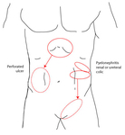
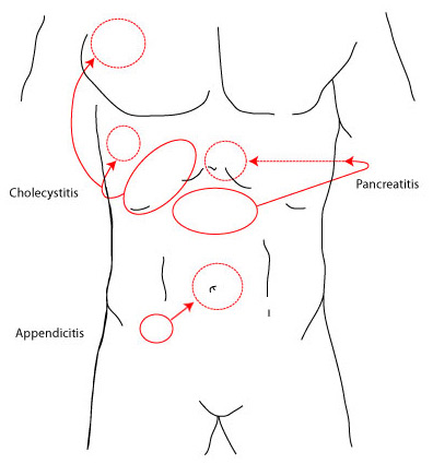
Solid circles represent the primary sites of pain and dotted circles represent the areas of referred pain
Created by BMJ Knowledge Centre
See this image in context in the following section/s:

Evaluation of acute abdomen
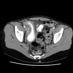
Ischemic bowel disease: CT angiogram: acute superior mesenteric artery thrombus
From the collection of Dr Jennifer Holder-Murray and Dr Alessandro Fichera
See this image in context in the following section/s:

Evaluation of acute abdomen
Solid circles represent the primary sites of pain and the dotted circles represent the areas of referred pain
Created by the BMJ Evidence Centre
See this image in context in the following section/s:

Evaluation of acute abdomen
Clostridium difficile-associated disease: CT scan of the abdomen showing gross thickening of the large bowel wall and obliteration of the lumen
Yates B, Murphy CM, et al. Pseudomembranous colitis in four patients with cystic fibrosis following lung transplantation. BMJ Case Reports. 2009; doi: 10.1136/bcr.11.2008.1218
See this image in context in the following section/s:

Evaluation of acute abdomen
Ischemic bowel disease: CT scan showing colonic thickening with pneumatosis intestinalis
From the collection of Dr Jennifer Holder-Murray and Dr Alessandro Fichera
See this image in context in the following section/s:
Evaluation of acute abdomen
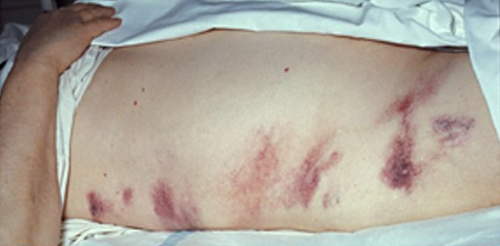
Grey-Turner sign (bruising of the flanks) in a 40-year-old woman with worsening epigastric pain of 5 days’ duration
Courtesy of Herbert L. Fred MD and Hendrik A. van Dijk
See this image in context in the following section/s:

Evaluation of acute abdomen
Intussusception: Site of intussusception as revealed by abdominal x-ray, showing the meniscus
From the collection of Dr David J. Hackam
See this image in context in the following section/s:
Evaluation of acute abdomen
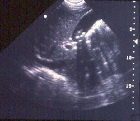
Intussusception: Transverse sonogram of the abdomen showing the donut sign (concentric rings within the lumen of a distended loop of bowel)
Adapted from the Student BMJ. 2008;16:76
See this image in context in the following section/s:

Evaluation of acute abdomen
Appendicitis: CT abdomen showing thickened appendix
Courtesy of Nasim Ahmed, MBBS, FACS
See this image in context in the following section/s:

Evaluation of acute abdomen
Ectopic pregnancy: Surgical extraction of ectopic pregnancy
From the personal collection of Dr Melissa Fries, Washington Hospital Center; used with permission
See this image in context in the following section/s:
Evaluation of acute abdomen
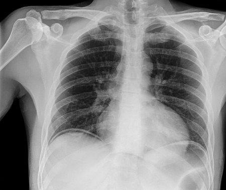
Abdominal free gas pockets, x-ray
Science Photo Library; used with permission
See this image in context in the following section/s:
Evaluation of acute abdomen
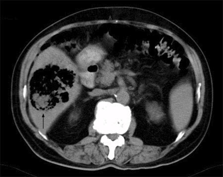
Liver abscess: A noncontrast abdominal CT scan showing a huge gas-containing liver abscess (arrow)
Adapted from BMJ Case Reports 2009 (doi:10.1136/bcr.08.2008.0638)
See this image in context in the following section/s:
Evaluation of acute abdomen
Intussusception: Ultrasound image showing invagination of a segment of bowel into the adjacent segment
BMJ Case Reports 2009; doi:10.1136/bcr.04.2009.1730
See this image in context in the following section/s:
Evaluation of acute abdomen
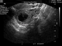
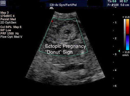
Ectopic pregnancy: Ultrasound image of ectopic pregnancy showing the donut sign
From the personal collection of Dr Melissa Fries, Washington Hospital Center; used with permission
See this image in context in the following section/s:

Evaluation of acute abdomen
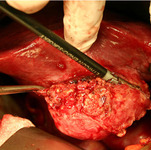
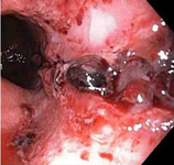
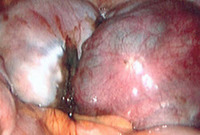
Cholecystitis: Operative photo showing acute cholecystitis
From the collection of Dr Charles Bellows
See this image in context in the following section/s:

Evaluation of acute abdomen
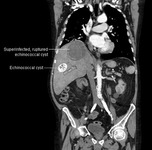
Liver abscess: CT scan showing 8 cm by 8 cm superinfected and ruptured echinococcal cyst, and a 4 cm by 4 cm echinococcal cyst in a 69-year-old man who presented with hypotension and chest pain radiating to the epigastric region
From the collection of MGH Massachusetts General Hospital radiology images
See this image in context in the following section/s:

Evaluation of acute abdomen
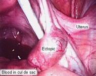
Ectopic pregnancy: Blood in cul de sac
From the personal collection of Dr Melissa Fries, Washington Hospital Center; used with permission
See this image in context in the following section/s:

Evaluation of acute abdomen
Crohn disease: Endoscopic view of normal terminal ileum
From the personal collection of Dr Charlotte Ford, North Middlesex Hospital Trust, London, UK
See this image in context in the following section/s:

Evaluation of acute abdomen
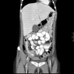
Crohn disease: CT scan demonstrating thickening of the terminal ileum in a patient with Crohn disease exacerbation
Provided by Drs Wissam Bleibel, Bishal Mainali, Chandrashekhar Thukral, and Mark A. Peppercorn
See this image in context in the following section/s:

Evaluation of acute abdomen
Endometriosis: Laparoscopic image of endometriotic nodule
From the collection of Dr Jonathon Solnik; used with permission
See this image in context in the following section/s:
Evaluation of acute abdomen
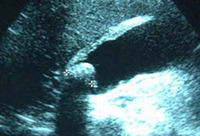
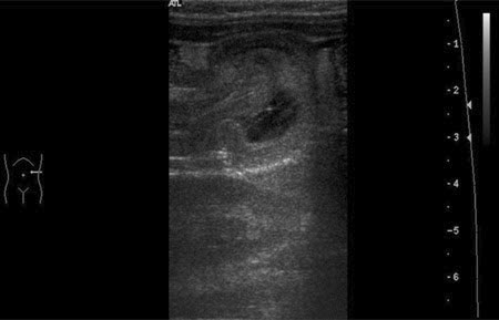
Gallbladder ultrasound demonstrating cholelithiasis with characteristic shadowing
Courtesy of Kuojen Tsao; used with permission
See this image in context in the following section/s:
Evaluation of acute abdomen
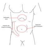
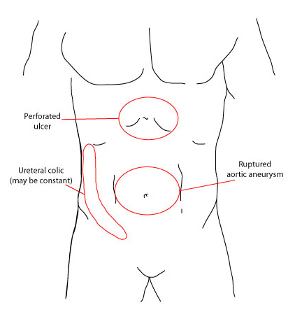
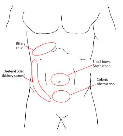
Areas of pain that present suddenly and severe in onset
Created by the BMJ Evidence Centre
See this image in context in the following section/s:
Evaluation of acute abdomen
Ectopic pregnancy: Ultrasound image of ectopic pregnancy
From the personal collection of Dr Melissa Fries, Washington Hospital Center; used with permission
See this image in context in the following section/s:

Evaluation of acute abdomen
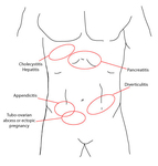
Areas of pain that present gradually or more progressively
Created by the BMJ Evidence Centre
See this image in context in the following section/s:
Evaluation of acute abdomen
Ectopic pregnancy: Blood in the abdomen
From the personal collection of Dr Melissa Fries, Washington Hospital Center; used with permission
See this image in context in the following section/s:
Evaluation of acute abdomen
Mallory-Weiss tear: Actively bleeding tear appears as a red longitudinal defect with normal surrounding mucosa
From the collection of Juan Carlos Munoz, MD, University of Florida
See this image in context in the following section/s:

Evaluation of acute abdomen
Intussusception: Abdominal x-ray showing impaired passage of barium at site of obstruction due to intussusception
From the collection of Dr David J. Hackam
See this image in context in the following section/s:
Evaluation of acute abdomen
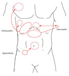
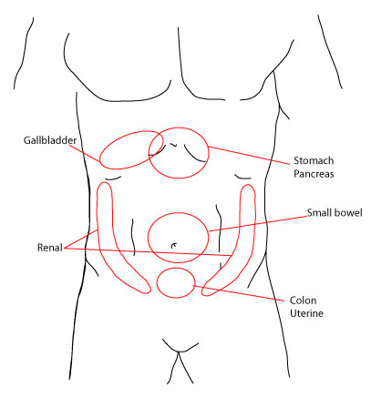
Common locations of visceral pain
Created by the BMJ Evidence Centre
See this image in context in the following section/s:

Evaluation of acute abdomen
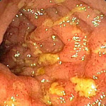
Crohn disease: Endoscopic view of Crohn ileitis
Provided by Drs Wissam Bleibel, Bishal Mainali, Chandrashekhar Thukral, and Mark A. Peppercorn
See this image in context in the following section/s:
Evaluation of acute abdomen
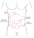
Areas of pain that present more colicky, crampy, and intermittent in nature
Created by the BMJ Evidence Centre
See this image in context in the following section/s:

Evaluation of acute abdomen
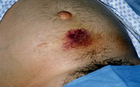
Cullen sign (periumbilical discoloration) in a 36-year-old man who presented with a 4-day history of severe epigastric pain following an alcoholic binge
Courtesy of Herbert L. Fred MD and Hendrik A van Dijk
See this image in context in the following section/s:
Evaluation of acute abdomen
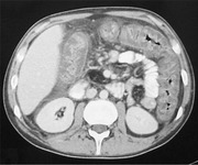
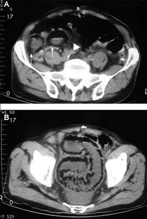
Ischemic bowel disease: 84-year-old man presenting with symptoms suggestive of ischemic bowel disease: (A) Abdominal CT revealing a massive circumferential and band-like air formation as intestinal pneumatosis (arrows) and pronounced edema of mesenteric fat (arrowhead) around necrotic bowel loops; (B) Another slice of abdominal CT showing long segmental pneumatosis of the small bowel
Lin I, Chang W, Shih S, et al. Bedside echogram in ischaemic bowel. BMJ Case Reports. 2009:bcr.2007.053462
See this image in context in the following section/s:

Evaluation of acute abdomen
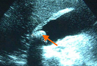
Ultrasound of acute cholecystitis and presence of gallstones: the arrow points to a gallstone in the fundus of the gallbladder with its echogenic shadow below
Courtesy of Charles Bellows and W. Scott Helton; used with permission
See this image in context in the following section/s:
Use of this content is subject to our disclaimer