Approach
Initial evaluation should include an extensive assessment of the patient's medical history and determination of the etiology. In patients not at risk for cirrhosis, other causes of ascites should be sought, such as cancer, congestive heart failure, tuberculosis, hemodialysis, or pancreatitis. Ascites due to more than one cause can be present in approximately 5% of patients, and the clinical picture can be confusing.[14] In patients with worsening ascites, noncompliance is a possible cause, along with liver disease decompensation due to development of hepatocellular carcinoma or superimposed acute on chronic liver injury.
History
Investigating risks for, and the presence of, liver disease is essential. The amount and duration of alcohol consumption should be reviewed in detail, as alcohol-related liver disease can cause ascites without cirrhosis. Ethanol ingestion of 80 g/day for 10 to 20 years is usually required for the development of cirrhosis, but lesser amounts may be sufficient for women and those with coexisting viral hepatitis.
A detailed history of the type of alcohol and amount consumed per day is helpful in calculating the total ethanol consumption. Alcohol content of beer is 3% to 8%, wine 7% to 18%, and spirits >37.5%. Standard alcoholic drinks in the US all contain about 0.6 fluid ounces of ethanol per glass (17.75 mL). A US standard drink is a 12-ounce can or bottle of beer, a 5-ounce glass of dinner wine, or a 1.5-ounce drink of 40% distilled spirits.
History of possible toxins or drugs such as acetaminophen should also be elicited.
Risk factors for hepatitis C should be assessed; these include blood transfusion before 1992, needle-sharing during intravenous drug misuse, shared use of a device for snorting of drugs, and tattoos. Hepatitis B should be considered in patients born in hyperendemic areas (i.e., Africa, sections of Asia, and Caribbean islands) and those with other risk factors such as multiple sexual partners, intravenous drug use, dialysis, healthcare workers, household contact with hepatitis B virus-infected people, or diagnosis of other STDs.
Family history of liver diseases such as hemochromatosis should be obtained. Joint pains and changes in skin color may be found in patients with hemochromatosis.
Metabolic dysfunction-associated steatohepatitis is a common cause of cirrhosis, with risk factors including obesity, diabetes, and hyperlipidemia.[29][30][31] Other causes of cirrhosis include autoimmune (e.g., autoimmune hepatitis, primary biliary cholangitis, primary sclerosing cholangitis) and metabolic liver diseases (e.g., hemochromatosis). A history of autoimmune diseases may be present in patients with autoimmune hepatitis. Patients with primary biliary cholangitis may complain of itching.
Shortness of breath on exertion or at rest with orthopnea suggests congestive heart failure. Patients may have a history of heart disease (e.g., coronary heart disease, prior heart surgery, chest pain on exertion, or percutaneous intervention). Patients with gastrointestinal malignancies may have a known malignancy or symptoms suggestive of malignant disease (e.g., loss of weight/appetite). Budd-Chiari syndrome presents with acute ascites and abdominal distension developing rapidly over days. A history of acute pancreatitis may be found in pancreatic ascites, and biliary ascites occurs following biliary surgery.
Immigrants from endemic areas for Schistosomiasis (South America, Africa) may have noncirrhotic portal hypertension secondary to a long disease course with spontaneous resolution (burned out Schistosomiasis).
Physical examination
Patients usually accumulate fluid rapidly, so they may experience early satiety and shortness of breath. The most useful physical finding in diagnosis is flank bulging and shifting dullness.[32] Assessment of conditions leading to ascites is important. Stigmata of cirrhosis such as vascular spiders, palmar erythema, and abdominal wall collaterals are useful, if present. Other signs of liver disease include jaundice, muscle wasting, leukonychia, and liver and spleen enlargement. Jugular venous distention suggests congestive heart failure or constrictive pericarditis in the absence of tense ascites, pulmonary hypertension, or renal insufficiency.
Assess for abdominal tenderness with rebound, which may indicate peritonitis. The sensitivity and specificity of physical examination can range from 50% to 94% and 29% to 82%, respectively, compared with ultrasound as reference standard.[32]
Some patients may develop hepatic hydrothorax (accumulation of fluid in the right pleural space). If a pleural effusion is present, chest examination typically reveals dullness to percussion, reduced or absent breath sounds, and lack of fremitus.[33] It may be possible to palpate ovarian mass on abdominal examination or bimanual examination. Patients with nephrotic syndrome would have prominent peripheral edema and history of kidney disease.
Imaging tests
In patients presenting with abdominal distention, ascites should first be differentiated from fat secondary to obesity. Ultrasound should be ordered if the physical finding is not definitive.[20][34] For diagnosis, approximately 1500 mL of ascitic fluid should be present for detection of flank dullness. The International Ascites Club has proposed the following ascites grading system:[35]
Grade 1: mild ascites detectable only by ultrasound
Grade 2: moderate ascites with moderate symmetrical distention of abdomen
Grade 3: large or gross ascites with marked abdominal distention.
Ultrasound of the abdomen is the most cost-effective modality to confirm ascites and to detect cirrhosis or malignancy. Doppler ultrasound studies may be useful for determining the patency of hepatic and portal veins and should be ordered in all patients. Portal vein thrombosis or stenosis of hepatic veins can be detected more accurately with Doppler ultrasound. A computed tomography scan or magnetic resonance imaging may be needed in some cases, such as suspected malignancy. A spleen diameter of more than 12 cm or recanalization of the umbilical veins indicates portal hypertension.[Figure caption and citation for the preceding image starts]: CT scan of abdomen showing massive ascites secondary to cirrhosis and hepatocellular carcinomaBrooklyn Hospital Center; used with permission [Citation ends].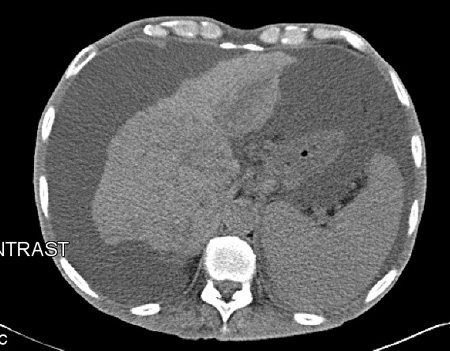
Routine laboratory tests
A basic metabolic profile and hepatic profile should be ordered in all patients. Spontaneous bacterial peritonitis is a risk factor for hepatorenal syndrome, and renal function should be monitored. Rising bilirubin and worsening coagulation profile suggest worsening cirrhosis. Calculating the Model for End-Stage Liver Disease (MELD) score and determining the Child-Pugh class would help in evaluation of underlying cirrhosis. The MELD score is based on objective measures (serum bilirubin, creatinine, sodium, and international normalized ratio); the Child-Pugh class is based on both objective and subjective measures (serum albumin, prothrombin time, serum bilirubin, degree of ascites, and encephalopathy).[36] [ Child Pugh classification for severity of liver disease Opens in new window ] Complete blood count is indicated to determine the platelet count, as thrombocytopenia is an indication of portal hypertension.
Serologic tests for hepatitis A, B, and C virus should be ordered routinely to determine the exposure status and need for immunization.
Abdominal paracentesis
A diagnostic abdominal paracentesis is indicated for patients with new-onset ascites upon any hospital admission, clinical deterioration (i.e., fever, abdominal pain or tenderness, mental status change, ileus, hypotension), gastrointestinal bleeding (high risk for spontaneous bacterial peritonitis), or any laboratory abnormality that indicates infection (i.e., peripheral leukocytosis, acidosis, worsening renal dysfunction).[20][37]
One study that used ultrasound to measure abdominal wall thickness and depth of ascites in 52 patients demonstrated that the left lower quadrant is the optimal site for paracentesis, due to a thinner abdominal wall and a greater depth of fluid.[38]
Postprocedural hemorrhage is very rare following diagnostic paracentesis.[39][40] Guidelines do not recommend routine prophylactic use of fresh frozen plasma or platelets before paracentesis.[41][42] Patients with disseminated intravascular coagulation or with clinical evidence of fibrinolysis should not undergo paracentesis until these conditions improve. Consult local guidance.
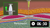
Demonstrates how to perform diagnostic and therapeutic abdominal paracentesis.
The routine tests for ascitic fluid are:[21]
Cell count and differential
Albumin
Total protein concentration
A simultaneous (or same day) serum albumin should be measured as well to calculate the serum ascites albumin gradient (SAAG).
If infection is suspected then ascitic fluid culture (in blood culture bottles) should be added to these routine tests.[21]
If the SAAG is <1.1 g/dL, suggesting a noncirrhotic cause of ascites, further tests on ascitic fluid may include:[21]
Measurement of glucose, lactate dehydrogenase, and amylase
Gram stain analysis
Tuberculosis smear and culture (if tuberculous peritonitis is suspected)
Cytology (for possible peritoneal carcinomatosis)
Adenosine deaminase (to distinguish between tuberculous peritonitis and carcinomatosis)
Brain natriuretic peptide (for underlying or additional cardiac disease)
Triglyceride concentration (if chylous ascites is suspected e.g., due to trauma or lymphatic obstruction)
Bilirubin (if bile ascites is suspected e.g., history of biliary surgery, abdominal trauma or cholecystitis)
Liver biopsy
When liver cirrhosis is suspected and clinical presentation is not very clear, liver biopsy may detect or rule out cirrhosis as the cause of ascites. Liver biopsy may also detect the etiology of cirrhosis such as hemochromatosis, alcohol-related liver disease, or viral hepatitis. However, characteristic features of the primary insult (e.g., metabolic dysfunction-associated steatotic liver disease [previously known as nonalcoholic fatty liver disease or autoimmune hepatitis]) may no longer be detectable by the time the procedure is carried out. Accurate staging of fibrosis and grading of inflammation can guide therapy in viral hepatitis.
Measurement of the hepatic venous pressure gradient (HVPG) can be achieved during transjugular liver biopsy. An HVPG ≥10 mmHg, indicating clinically significant portal hypertension, may help in differentiating etiology of ascites.[48]
Use of this content is subject to our disclaimer